Differential Reactivity Between the Two Copper Sites of Peptidylglycine alpha-Hydroxylating Monooxygenase (PHM)
Chufan, E.E., Prigge, S.T., Siebert, X., Eipper, B.A., Mains, R.E., Amzel, L.M.(2010) J Am Chem Soc 132: 15565-15572
- PubMed: 20958070
- DOI: https://doi.org/10.1021/ja103117r
- Primary Citation of Related Structures:
3MIB, 3MIC, 3MID, 3MIE, 3MIF, 3MIG, 3MIH, 3MLJ, 3MLK, 3MLL - PubMed Abstract:
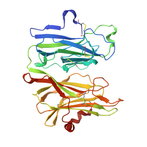
Peptidylglycine α-hydroxylating monooxygenase (PHM) catalyzes the stereospecific hydroxylation of the Cα of C-terminal glycine-extended peptides and proteins, the first step in the activation of many peptide hormones, growth factors, and neurotransmitters. The crystal structure of the enzyme revealed two nonequivalent Cu sites (Cu(M) and Cu(H)) separated by ∼11 Å. In the resting state of the enzyme, Cu(M) is coordinated in a distorted tetrahedral geometry by one methionine, two histidines, and a water molecule. The coordination site of the water molecule is the position where external ligands bind. The Cu(H) has a planar T-shaped geometry with three histidines residues and a vacant position that could potentially be occupied by a fourth ligand. Although the catalytic mechanism of PHM and the role of the metals are still being debated, Cu(M) is identified as the metal involved in catalysis, while Cu(H) is associated with electron transfer. To further probe the role of the metals, we studied how small molecules such as nitrite (NO(2)(-)), azide (N(3)(-)), and carbon monoxide (CO) interact with the PHM copper ions. The crystal structure of an oxidized nitrite-soaked PHMcc, obtained by soaking for 20 h in mother liquor supplemented with 300 mM NaNO(2), shows that nitrite anion coordinates Cu(M) in an asymmetric bidentate fashion. Surprisingly, nitrite does not bind Cu(H), despite the high concentration used in the experiments (nitrite/protein > 1000). Similarly, azide and carbon monoxide coordinate Cu(M) but not Cu(H) in the PHMcc crystal structures obtained by cocrystallization with 40 mM NaN(3) and by soaking CO under 3 atm of pressure for 30 min. This lack of reactivity at the Cu(H) is also observed in the reduced form of the enzyme: CO binds Cu(M) but not Cu(H) in the structure of PHMcc obtained by exposure of a crystal to 3 atm CO for 15 min in the presence of 5 mM ascorbic acid (reductant). The necessity of Cu(H) to maintain its redox potential in a narrow range compatible with its role as an electron-transfer site seems to explain the lack of coordination of small molecules to Cu(H); coordination of any external ligand will certainly modify its redox potential.
- Department of Biophysics and Biophysical Chemistry, Johns Hopkins School of Medicine, and Molecular Microbiology and Immunology, Johns Hopkins University, Baltimore, Maryland 21205, United States.
Organizational Affiliation:


