3MG5 | pdb_00003mg5

Core-streptavidin mutant F130L in complex with biotin


Experimental Data Snapshot

  • Method: X-RAY DIFFRACTION
  • Resolution: 1.30 Å
  • R-Value Free: 
    0.178 (Depositor), 0.179 (DCC) 
  • R-Value Work: 
    0.132 (Depositor), 0.140 (DCC) 
  • R-Value Observed: 
    0.134 (Depositor) 

Starting Model: experimental
View more details

wwPDB Validation 3D Report Full Report

Validation slider image for 3MG5

Ligand Structure Quality Assessment 


This is version 1.4 of the entry. See complete history

Literature

A distal point mutation in the streptavidin-biotin complex preserves structure but diminishes binding affinity: experimental evidence of electronic polarization effects?

Baugh, L.Le Trong, I.Cerutti, D.S.Gulich, S.Stayton, P.S.Stenkamp, R.E.Lybrand, T.P.

(2010) Biochemistry 49: 4568-4570

  • DOI: https://doi.org/10.1021/bi1005392
  • Primary Citation Related Structures: 
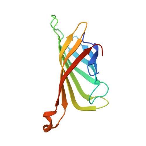
    3MG5

  • PubMed Abstract: 

    We have identified a distal point mutation in streptavidin that causes a 1000-fold reduction in biotin binding affinity without disrupting the equilibrium complex structure. The F130L mutation creates a small cavity occupied by a water molecule; however, all neighboring side chain positions are preserved, and protein-biotin hydrogen bonds are unperturbed. Molecular dynamics simulations reveal a reduced mobility of biotin binding residues but no observable destabilization of protein-ligand interactions. Our combined structural and computational studies suggest that the additional water molecule may affect binding affinity through an electronic polarization effect that impacts the highly cooperative hydrogen bonding network in the biotin binding pocket.


  • Organizational Affiliation
    • Department of Bioengineering, University of Washington, Seattle, Washington 98195, USA.

Macromolecule Content 

  • Total Structure Weight: 54.98 kDa 
  • Atom Count: 4,315 
  • Modeled Residue Count: 479 
  • Deposited Residue Count: 508 
  • Unique protein chains: 1

Macromolecules

Find similar proteins by:|  3D Structure
Entity ID: 1
MoleculeChains  Sequence LengthOrganismDetailsImage
Streptavidin
A, B, C, D
127Streptomyces avidiniiMutation(s): 1 
UniProt
Find proteins for P22629 (Streptomyces avidinii)
Explore P22629 
Go to UniProtKB:  P22629
Entity Groups
Sequence Clusters30% Identity50% Identity70% Identity90% Identity95% Identity100% Identity
UniProt GroupP22629
Sequence Annotations
Expand
Reference Sequence

Small Molecules

Ligands 2 Unique
IDChains Name / Formula / InChI Key2D Diagram3D Interactions
BTN

Query on BTN


Download:Ideal Coordinates CCD File
E [auth A],
H [auth B],
L [auth C],
P [auth D]
BIOTIN
C10 H16 N2 O3 S
YBJHBAHKTGYVGT-ZKWXMUAHSA-N
GOL

Query on GOL


Download:Ideal Coordinates CCD File
F [auth A]
G [auth A]
I [auth B]
J [auth B]
K [auth B]
F [auth A],
G [auth A],
I [auth B],
J [auth B],
K [auth B],
M [auth C],
N [auth C],
O [auth C],
Q [auth D],
R [auth D],
S [auth D]
GLYCEROL
C3 H8 O3
PEDCQBHIVMGVHV-UHFFFAOYSA-N

Experimental Data & Validation

Experimental Data

  • Method: X-RAY DIFFRACTION
  • Resolution: 1.30 Å
  • R-Value Free:  0.178 (Depositor), 0.179 (DCC) 
  • R-Value Work:  0.132 (Depositor), 0.140 (DCC) 
  • R-Value Observed: 0.134 (Depositor) 
Space Group: P 1 21 1
Unit Cell:
Length ( Å )Angle ( ˚ )
a = 50.91α = 90
b = 97.53β = 112.3
c = 52.68γ = 90
Software Package:
Software NamePurpose
CCP4model building
REFMACrefinement
XDSdata reduction
XDSdata scaling
CCP4phasing

Structure Validation

View Full Validation Report


Ligand Structure Quality Assessment 


Entry History 

Deposition Data

Revision History  (Full details and data files)

  • Version 1.0: 2010-05-26
    Type: Initial release
  • Version 1.1: 2011-07-13
    Changes: Version format compliance
  • Version 1.2: 2017-11-08
    Changes: Refinement description
  • Version 1.3: 2021-10-06
    Changes: Database references, Derived calculations
  • Version 1.4: 2023-09-06
    Changes: Data collection, Refinement description