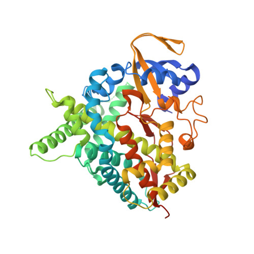
3MDR

Tranylcypromine complex of Cytochrome P450 46A1


Experimental Data Snapshot

  • Method: X-RAY DIFFRACTION
  • Resolution: 2.00 Å
  • R-Value Free: 0.244 
  • R-Value Work: 0.225 
  • R-Value Observed: 0.226 

Starting Model: experimental
View more details

wwPDB Validation   3D Report Full Report


Ligand Structure Quality Assessment 


This is version 1.3 of the entry. See complete history


Literature

Structural basis of drug binding to CYP46A1, an enzyme that controls cholesterol turnover in the brain.

Mast, N.Charvet, C.Pikuleva, I.A.Stout, C.D.

(2010) J Biol Chem 285: 31783-31795

  • DOI: https://doi.org/10.1074/jbc.M110.143313
  • Primary Citation of Related Structures:  
    3MDM, 3MDR, 3MDT, 3MDV

  • PubMed Abstract: 

    Cytochrome P450 46A1 (CYP46A1) initiates the major pathway of cholesterol elimination from the brain and thereby controls cholesterol turnover in this organ. We determined x-ray crystal structures of CYP46A1 in complex with four structurally distinct pharmaceuticals; antidepressant tranylcypromine (2.15 Å), anticonvulsant thioperamide (1.65 Å), antifungal voriconazole (2.35 Å), and antifungal clotrimazole (2.50 Å). All four drugs are nitrogen-containing compounds that have nanomolar affinity for CYP46A1 in vitro yet differ in size, shape, hydrophobicity, and type of the nitrogen ligand. Structures of the co-complexes demonstrate that each drug binds in a single orientation to the active site with tranylcypromine, thioperamide, and voriconazole coordinating the heme iron via their nitrogen atoms and clotrimazole being at a 4 Å distance from the heme iron. We show here that clotrimazole is also a substrate for CYP46A1. High affinity for CYP46A1 is determined by a set of specific interactions, some of which were further investigated by solution studies using structural analogs of the drugs and the T306A CYP46A1 mutant. Collectively, our results reveal how diverse inhibitors can be accommodated in the CYP46A1 active site and provide an explanation for the observed differences in the drug-induced spectral response. Co-complexes with tranylcypromine, thioperamide, and voriconazole represent the first structural characterization of the drug binding to a P450 enzyme.


  • Organizational Affiliation

    Department of Ophthalmology and Visual Sciences, Case Western Reserve University, Cleveland, Ohio 44106, USA.


Macromolecules
Find similar proteins by:  (by identity cutoff)  |  3D Structure
Entity ID: 1
MoleculeChains Sequence LengthOrganismDetailsImage
Cholesterol 24-hydroxylase
A, B
456Homo sapiensMutation(s): 0 
Gene Names: CYP46CYP46A1
EC: 1.14.13.98 (PDB Primary Data), 1.14.14.25 (UniProt)
UniProt & NIH Common Fund Data Resources
Find proteins for Q9Y6A2 (Homo sapiens)
Explore Q9Y6A2 
Go to UniProtKB:  Q9Y6A2
PHAROS:  Q9Y6A2
GTEx:  ENSG00000036530 
Entity Groups  
Sequence Clusters30% Identity50% Identity70% Identity90% Identity95% Identity100% Identity
UniProt GroupQ9Y6A2
Sequence Annotations
Expand
  • Reference Sequence
Experimental Data & Validation

Experimental Data

  • Method: X-RAY DIFFRACTION
  • Resolution: 2.00 Å
  • R-Value Free: 0.244 
  • R-Value Work: 0.225 
  • R-Value Observed: 0.226 
  • Space Group: I 41
Unit Cell:
Length ( Å )Angle ( ˚ )
a = 121.19α = 90
b = 121.19β = 90
c = 143.99γ = 90
Software Package:
Software NamePurpose
REFMACrefinement
CNSrefinement
PDB_EXTRACTdata extraction
Blu-Icedata collection
MOSFLMdata reduction
SCALAdata scaling
MOLREPphasing
SCALAdata processing

Structure Validation

View Full Validation Report


Ligand Structure Quality Assessment 


Entry History 

Deposition Data

Revision History  (Full details and data files)

  • Version 1.0: 2010-07-28
    Type: Initial release
  • Version 1.1: 2011-07-13
    Changes: Version format compliance
  • Version 1.2: 2019-07-17
    Changes: Data collection, Refinement description
  • Version 1.3: 2023-09-06
    Changes: Data collection, Database references, Derived calculations, Refinement description