3MAY | pdb_00003may

Crystal structure of a secreted Mycobacterium tuberculosis heme-binding protein


Experimental Data Snapshot

  • Method: X-RAY DIFFRACTION
  • Resolution: 2.50 Å
  • R-Value Free: 
    0.256 (Depositor), 0.231 (DCC) 
  • R-Value Work: 
    0.195 (Depositor), 0.202 (DCC) 
  • R-Value Observed: 
    0.198 (Depositor) 

wwPDB Validation 3D Report Full Report

Validation slider image for 3MAY

This is version 1.2 of the entry. See complete history

Literature

Discovery and characterization of a unique mycobacterial heme acquisition system.

Tullius, M.V.Harmston, C.A.Owens, C.P.Chim, N.Morse, R.P.McMath, L.M.Iniguez, A.Kimmey, J.M.Sawaya, M.R.Whitelegge, J.P.Horwitz, M.A.Goulding, C.W.

(2011) Proc Natl Acad Sci U S A 108: 5051-5056

  • DOI: https://doi.org/10.1073/pnas.1009516108
  • Primary Citation Related Structures: 
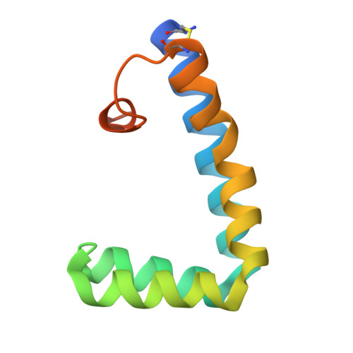
    3MAY

  • PubMed Abstract: 

    Mycobacterium tuberculosis must import iron from its host for survival, and its siderophore-dependent iron acquisition pathways are well established. Here we demonstrate a newly characterized pathway, whereby M. tuberculosis can use free heme and heme from hemoglobin as an iron source. Significantly, we identified the genomic region, Rv0202c-Rv0207c, responsible for the passage of heme iron across the mycobacterial membrane. Key players of this heme uptake system were characterized including a secreted protein and two transmembrane proteins, all three specific to mycobacteria. Furthermore, the crystal structure of the key heme carrier protein Rv0203 was found to have a unique fold. The discovery of a unique mycobacterial heme acquisition pathway opens new avenues of exploration into mycobacterial therapeutics.


  • Organizational Affiliation
    • Departments of Molecular Biology and Biochemistry, University of California, Irvine, CA 92697, USA.

Macromolecule Content 

  • Total Structure Weight: 83.28 kDa 
  • Atom Count: 5,379 
  • Modeled Residue Count: 743 
  • Deposited Residue Count: 808 
  • Unique protein chains: 1

Macromolecules

Find similar proteins by:|  3D Structure
Entity ID: 1
MoleculeChains  Sequence LengthOrganismDetailsImage
POSSIBLE EXPORTED PROTEIN101Mycobacterium tuberculosisMutation(s): 0 
Gene Names: MT0213Rv0203
UniProt
Find proteins for O53654 (Mycobacterium tuberculosis (strain CDC 1551 / Oshkosh))
Explore O53654 
Go to UniProtKB:  O53654
Entity Groups
Sequence Clusters30% Identity50% Identity70% Identity90% Identity95% Identity100% Identity
UniProt GroupO53654
Sequence Annotations
Expand
Reference Sequence

Experimental Data & Validation

Experimental Data

  • Method: X-RAY DIFFRACTION
  • Resolution: 2.50 Å
  • R-Value Free:  0.256 (Depositor), 0.231 (DCC) 
  • R-Value Work:  0.195 (Depositor), 0.202 (DCC) 
  • R-Value Observed: 0.198 (Depositor) 
Space Group: P 41
Unit Cell:
Length ( Å )Angle ( ˚ )
a = 72.671α = 90
b = 72.671β = 90
c = 173.909γ = 90
Software Package:
Software NamePurpose
HKL-2000data collection
MLPHAREphasing
REFMACrefinement
HKL-2000data reduction
SCALEPACKdata scaling

Structure Validation

View Full Validation Report


Entry History 

Deposition Data

Revision History  (Full details and data files)

  • Version 1.0: 2011-03-23
    Type: Initial release
  • Version 1.1: 2011-07-13
    Changes: Version format compliance
  • Version 1.2: 2024-11-06
    Changes: Data collection, Database references, Structure summary