


Crystal Structure of Phosphoribosylaminoimidazole Synthetase from Francisella tularensis
Maltseva, N., Kim, Y., Hasseman, J., Anderson, W.F., Joachimiak, A.To be published.
Experimental Data Snapshot
Entity ID: 1 | |||||
|---|---|---|---|---|---|
| Molecule | Chains | Sequence Length | Organism | Details | Image |
| Phosphoribosylformylglycinamidine cyclo-ligase | 350 | Francisella tularensis subsp. tularensis SCHU S4 | Mutation(s): 0 Gene Names: purM EC: 6.3.3.1 | 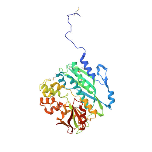 | |
UniProt | |||||
Entity Groups | |||||
| Sequence Clusters | 30% Identity50% Identity70% Identity90% Identity95% Identity100% Identity | ||||
| UniProt Group | Q5NGF2 | ||||
Sequence AnnotationsExpand | |||||
Reference Sequence | |||||
| Ligands 5 Unique | |||||
|---|---|---|---|---|---|
| ID | Chains | Name / Formula / InChI Key | 2D Diagram | 3D Interactions | |
| AMP Download:Ideal Coordinates CCD File | F [auth A] | ADENOSINE MONOPHOSPHATE C10 H14 N5 O7 P UDMBCSSLTHHNCD-KQYNXXCUSA-N |  | ||
| TRS Download:Ideal Coordinates CCD File | V [auth B] | 2-AMINO-2-HYDROXYMETHYL-PROPANE-1,3-DIOL C4 H12 N O3 LENZDBCJOHFCAS-UHFFFAOYSA-O |  | ||
| SO4 Download:Ideal Coordinates CCD File | C [auth A] D [auth A] E [auth A] G [auth A] K [auth A] | SULFATE ION O4 S QAOWNCQODCNURD-UHFFFAOYSA-L |  | ||
| ACY Download:Ideal Coordinates CCD File | H [auth A], J [auth A], U [auth B] | ACETIC ACID C2 H4 O2 QTBSBXVTEAMEQO-UHFFFAOYSA-N |  | ||
| FMT Download:Ideal Coordinates CCD File | I [auth A], M [auth B] | FORMIC ACID C H2 O2 BDAGIHXWWSANSR-UHFFFAOYSA-N |  | ||
| Modified Residues 1 Unique | |||||
|---|---|---|---|---|---|
| ID | Chains | Type | Formula | 2D Diagram | Parent |
| MSE Query on MSE | A, B | L-PEPTIDE LINKING | C5 H11 N O2 Se |  | MET |
| Length ( Å ) | Angle ( ˚ ) |
|---|---|
| a = 62.708 | α = 90 |
| b = 90.56 | β = 90 |
| c = 118.989 | γ = 90 |
| Software Name | Purpose |
|---|---|
| SBC-Collect | data collection |
| HKL-3000 | data collection |
| HKL-3000 | phasing |
| SHELX | model building |
| MLPHARE | phasing |
| DM | model building |
| SOLVE | phasing |
| RESOLVE | model building |
| PHENIX | refinement |
| HKL-3000 | data reduction |
| HKL-3000 | data scaling |
| SHELX | phasing |
| DM | phasing |
| RESOLVE | phasing |


