3M2C | pdb_00003m2c

Crystallographic and Single Crystal Spectral Analysis of the Peroxidase Ferryl Intermediate


Experimental Data Snapshot

  • Method: X-RAY DIFFRACTION
  • Resolution: 1.40 Å
  • R-Value Work: 
    0.116 (Depositor), 0.140 (DCC) 

wwPDB Validation   3D Report Full Report


Ligand Structure Quality Assessment 

Created with Raphaël 2.3.0Worse 01 BetterLigand structure goodness of fit to experimental dataBest fitted HEMClick on this verticalbar to view details

This is version 1.3 of the entry. See complete history


Literature

Crystallographic and single-crystal spectral analysis of the peroxidase ferryl intermediate.

Meharenna, Y.T.Doukov, T.Li, H.Soltis, S.M.Poulos, T.L.

(2010) Biochemistry 49: 2984-2986

  • DOI: https://doi.org/10.1021/bi100238r
  • Primary Citation of Related Structures:  
    3M23, 3M25, 3M26, 3M27, 3M28, 3M29, 3M2A, 3M2B, 3M2C, 3M2D, 3M2E, 3M2F, 3M2G, 3M2H, 3M2I

  • PubMed Abstract: 

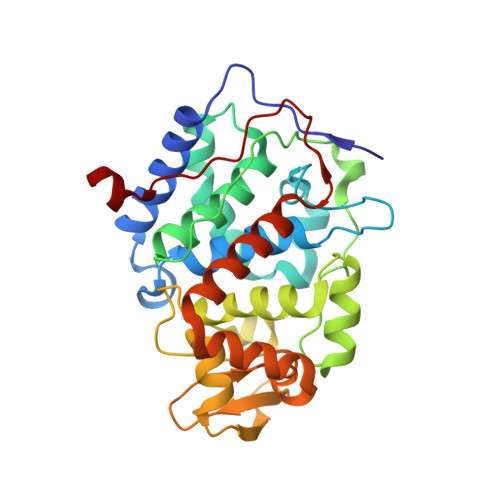
    The ferryl [Fe(IV)O] intermediate is important in many heme enzymes, and thus, the precise nature of the Fe(IV)-O bond is critical in understanding enzymatic mechanisms. The 1.40 A crystal structure of cytochrome c peroxidase Compound I has been determined as a function of X-ray dose while the visible spectrum was being monitored. The Fe-O bond increases in length from 1.73 A in the low-X-ray dose structure to 1.90 A in the high-dose structure. The low-dose structure correlates well with an Fe(IV) horizontal lineO bond, while we postulate that the high-dose structure is the cryo-trapped Fe(III)-OH species previously thought to be an Fe(IV)-OH species.


  • Organizational Affiliation

    Department of Molecular Biology and Biochemistry, University of California, Irvine, California 92697-3900, USA.


Macromolecules
Find similar proteins by:  (by identity cutoff)  |  3D Structure
Entity ID: 1
MoleculeChains Sequence LengthOrganismDetailsImage
Cytochrome c peroxidase, mitochondrial291Saccharomyces cerevisiaeMutation(s): 1 
Gene Names: CCPCCP1CPOYKR066C
EC: 1.11.1.5
UniProt
Find proteins for P00431 (Saccharomyces cerevisiae (strain ATCC 204508 / S288c))
Explore P00431 
Go to UniProtKB:  P00431
Entity Groups  
Sequence Clusters30% Identity50% Identity70% Identity90% Identity95% Identity100% Identity
UniProt GroupP00431
Sequence Annotations
Expand
  • Reference Sequence
Experimental Data & Validation

Experimental Data

  • Method: X-RAY DIFFRACTION
  • Resolution: 1.40 Å
  • R-Value Work:  0.116 (Depositor), 0.140 (DCC) 
Space Group: P 21 21 21
Unit Cell:
Length ( Å )Angle ( ˚ )
a = 107.019α = 90
b = 74.721β = 90
c = 50.857γ = 90
Software Package:
Software NamePurpose
MOSFLMdata reduction
SHELXmodel building
SHELXL-97refinement
HKL-2000data scaling
SHELXphasing

Structure Validation

View Full Validation Report


Ligand Structure Quality Assessment 

Created with Raphaël 2.3.0Worse 01 BetterLigand structure goodness of fit to experimental dataBest fitted HEMClick on this verticalbar to view details

Entry History 

Deposition Data

Revision History  (Full details and data files)

  • Version 1.0: 2010-05-12
    Type: Initial release
  • Version 1.1: 2011-07-13
    Changes: Version format compliance
  • Version 1.2: 2021-10-13
    Changes: Database references, Derived calculations
  • Version 1.3: 2024-02-21
    Changes: Data collection