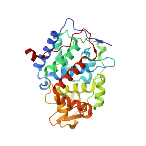
Crystallographic and single-crystal spectral analysis of the peroxidase ferryl intermediate.
Meharenna, Y.T., Doukov, T., Li, H., Soltis, S.M., Poulos, T.L.(2010) Biochemistry 49: 2984-2986
- PubMed: 20230048
- DOI: https://doi.org/10.1021/bi100238r
- Primary Citation of Related Structures:
3M23, 3M25, 3M26, 3M27, 3M28, 3M29, 3M2A, 3M2B, 3M2C, 3M2D, 3M2E, 3M2F, 3M2G, 3M2H, 3M2I - PubMed Abstract:
The ferryl [Fe(IV)O] intermediate is important in many heme enzymes, and thus, the precise nature of the Fe(IV)-O bond is critical in understanding enzymatic mechanisms. The 1.40 A crystal structure of cytochrome c peroxidase Compound I has been determined as a function of X-ray dose while the visible spectrum was being monitored. The Fe-O bond increases in length from 1.73 A in the low-X-ray dose structure to 1.90 A in the high-dose structure. The low-dose structure correlates well with an Fe(IV) horizontal lineO bond, while we postulate that the high-dose structure is the cryo-trapped Fe(III)-OH species previously thought to be an Fe(IV)-OH species.
Organizational Affiliation:
Department of Molecular Biology and Biochemistry, University of California, Irvine, California 92697-3900, USA.


