3M1J | pdb_00003m1j

The crystal structure of a NAMI A-Carbonic Anhydrase II adduct discloses the mode of action of this novel anticancer metallodrug


Experimental Data Snapshot

  • Method: X-RAY DIFFRACTION
  • Resolution: 1.80 Å
  • R-Value Free: 
    0.235 (Depositor), 0.194 (DCC) 
  • R-Value Work: 
    0.193 (Depositor), 0.194 (DCC) 
  • R-Value Observed: 
    0.195 (Depositor) 

Starting Model: experimental
View more details

wwPDB Validation 3D Report Full Report

Validation slider image for 3M1J

Ligand Structure Quality Assessment 


This is version 1.3 of the entry. See complete history

Literature

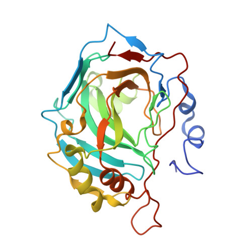
The x-ray structure of the adduct between NAMI-A and carbonic anhydrase provides insights into the reactivity of this metallodrug with proteins

Casini, A.Temperini, C.Gabbiani, C.Supuran, C.T.Messori, L.

(2010) ChemMedChem 5: 1989-1994

Macromolecule Content 

  • Total Structure Weight: 29.92 kDa 
  • Atom Count: 2,290 
  • Modeled Residue Count: 257 
  • Deposited Residue Count: 260 
  • Unique protein chains: 1

Macromolecules

Find similar proteins by:|  3D Structure
Entity ID: 1
MoleculeChains  Sequence LengthOrganismDetailsImage
Carbonic anhydrase 2260Homo sapiensMutation(s): 0 
Gene Names: CA2
EC: 4.2.1.1 (PDB Primary Data), 4.2.1.69 (UniProt)
UniProt & NIH Common Fund Data Resources
Find proteins for P00918 (Homo sapiens)
Explore P00918 
Go to UniProtKB:  P00918
PHAROS:  P00918
GTEx:  ENSG00000104267 
Entity Groups
Sequence Clusters30% Identity50% Identity70% Identity90% Identity95% Identity100% Identity
UniProt GroupP00918
Sequence Annotations
Expand
Reference Sequence

Small Molecules

Ligands 5 Unique
IDChains Name / Formula / InChI Key2D Diagram3D Interactions
MBO

Query on MBO


Download:Ideal Coordinates CCD File
D [auth A]MERCURIBENZOIC ACID
C7 H5 Hg O2
FVFZSVRSDNUCGG-UHFFFAOYSA-N
RU

Query on RU


Download:Ideal Coordinates CCD File
C [auth A]RUTHENIUM ION
Ru
BPEVHDGLPIIAGH-UHFFFAOYSA-N
DMS

Query on DMS


Download:Ideal Coordinates CCD File
E [auth A]DIMETHYL SULFOXIDE
C2 H6 O S
IAZDPXIOMUYVGZ-UHFFFAOYSA-N
IMD

Query on IMD


Download:Ideal Coordinates CCD File
F [auth A]IMIDAZOLE
C3 H5 N2
RAXXELZNTBOGNW-UHFFFAOYSA-O
ZN

Query on ZN


Download:Ideal Coordinates CCD File
B [auth A]ZINC ION
Zn
PTFCDOFLOPIGGS-UHFFFAOYSA-N

Experimental Data & Validation

Experimental Data

  • Method: X-RAY DIFFRACTION
  • Resolution: 1.80 Å
  • R-Value Free:  0.235 (Depositor), 0.194 (DCC) 
  • R-Value Work:  0.193 (Depositor), 0.194 (DCC) 
  • R-Value Observed: 0.195 (Depositor) 
Space Group: P 1 21 1
Unit Cell:
Length ( Å )Angle ( ˚ )
a = 42.1α = 90
b = 41.55β = 104.51
c = 72.29γ = 90
Software Package:
Software NamePurpose
CrysalisProdata collection
AMoREphasing
REFMACrefinement
CrysalisProdata reduction
SCALEPACKdata scaling

Structure Validation

View Full Validation Report


Ligand Structure Quality Assessment 


Entry History 

Deposition Data

Revision History  (Full details and data files)

  • Version 1.0: 2011-01-19
    Type: Initial release
  • Version 1.1: 2011-07-13
    Changes: Version format compliance
  • Version 1.2: 2017-11-08
    Changes: Refinement description
  • Version 1.3: 2023-11-01
    Changes: Data collection, Database references, Derived calculations, Refinement description