3LYN | pdb_00003lyn

STRUCTURE OF GREEN ABALONE LYSIN DIMER


Experimental Data Snapshot

  • Method: X-RAY DIFFRACTION
  • Resolution: 1.70 Å
  • R-Value Free: 
    0.259 (Depositor) 
  • R-Value Work: 
    0.203 (DCC) 
  • R-Value Observed: 
    0.213 (Depositor) 

wwPDB Validation 3D Report Full Report

Validation slider image for 3LYN

This is version 1.4 of the entry. See complete history

Literature

The high resolution crystal structure of green abalone sperm lysin: implications for species-specific binding of the egg receptor.

Kresge, N.Vacquier, V.D.Stout, C.D.

(2000) J Mol Biology 296: 1225-1234

  • DOI: https://doi.org/10.1006/jmbi.2000.3533
  • Primary Citation Related Structures: 
    3LYN

  • PubMed Abstract: 

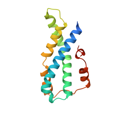
    Abalone sperm lysin is a 16 kDa acrosomal protein used by sperm to create a hole in the egg vitelline envelope. Lysins from seven California abalone exhibit species-specificity in binding to their egg receptor, and range in sequence identity from 63 % to 90 %. The crystal structure of the sperm lysin dimer from Haliotis fulgens (green abalone) has been determined to 1.71 A by multiple isomorphous replacement. Comparisons with the structure of the lysin dimer from Haliotis rufescens (red abalone) reveal a similar overall fold and conservation of features contributing to lysin's amphipathic character. The two structures do, however, exhibit differences in surface residues and electrostatics. A large clustering of non-conserved surface residues around the waist and clefts of the dimer, and differences in charged residues around these regions, indicate areas of the molecule which may be involved in species-specific egg recognition.


  • Organizational Affiliation
    • Department of Molecular Biology, The Scripps Research Institute, La Jolla, CA 92037-1093, USA.

Macromolecule Content 

  • Total Structure Weight: 32.69 kDa 
  • Atom Count: 2,271 
  • Modeled Residue Count: 246 
  • Deposited Residue Count: 272 
  • Unique protein chains: 1

Macromolecules

Find similar proteins by:|  3D Structure
Entity ID: 1
MoleculeChains  Sequence LengthOrganismDetailsImage
SPERM LYSIN
A, B
136Haliotis fulgensMutation(s): 0 
UniProt
Find proteins for Q01381 (Haliotis fulgens)
Explore Q01381 
Go to UniProtKB:  Q01381
Entity Groups
Sequence Clusters30% Identity50% Identity70% Identity90% Identity95% Identity100% Identity
UniProt GroupQ01381
Sequence Annotations
Expand
Reference Sequence

Experimental Data & Validation

Experimental Data

  • Method: X-RAY DIFFRACTION
  • Resolution: 1.70 Å
  • R-Value Free:  0.259 (Depositor) 
  • R-Value Work:  0.203 (DCC) 
  • R-Value Observed: 0.213 (Depositor) 
Space Group: P 61
Unit Cell:
Length ( Å )Angle ( ˚ )
a = 119.17α = 90
b = 119.17β = 90
c = 51.29γ = 120
Software Package:
Software NamePurpose
SOLVEphasing
SHARPphasing
SHELXLrefinement
MOSFLMdata reduction
CCP4data scaling

Structure Validation

View Full Validation Report


Entry History 

Deposition Data

Revision History  (Full details and data files)

  • Version 1.0: 2000-03-15
    Type: Initial release
  • Version 1.1: 2008-04-26
    Changes: Version format compliance
  • Version 1.2: 2011-07-13
    Changes: Version format compliance
  • Version 1.3: 2017-10-04
    Changes: Refinement description
  • Version 1.4: 2023-12-27
    Changes: Data collection, Database references