


Crystal structure of Putative histidinol-phosphate aminotransferase (YP_050345.1) from Erwinia carotovora atroseptica SCRI1043 at 1.80 A resolution
Joint Center for Structural Genomics (JCSG)To be published.
Experimental Data Snapshot
Entity ID: 1 | |||||
|---|---|---|---|---|---|
| Molecule | Chains | Sequence Length | Organism | Details | Image |
| Putative histidinol-phosphate aminotransferase | 354 | Pectobacterium atrosepticum | Mutation(s): 0 Gene Names: ECA2250 | 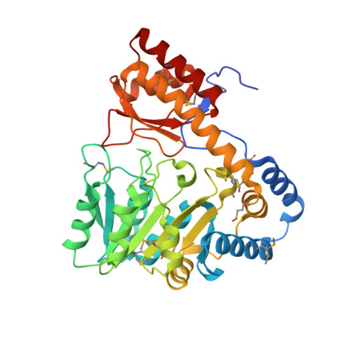 | |
UniProt | |||||
Entity Groups | |||||
| Sequence Clusters | 30% Identity50% Identity70% Identity90% Identity95% Identity100% Identity | ||||
| UniProt Group | Q6D4Z0 | ||||
Sequence AnnotationsExpand | |||||
Reference Sequence | |||||
| Ligands 3 Unique | |||||
|---|---|---|---|---|---|
| ID | Chains | Name / Formula / InChI Key | 2D Diagram | 3D Interactions | |
| PLP Download:Ideal Coordinates CCD File | BA [auth D], E [auth A], M [auth B], R [auth C] | PYRIDOXAL-5'-PHOSPHATE C8 H10 N O6 P NGVDGCNFYWLIFO-UHFFFAOYSA-N |  | ||
| CIT Download:Ideal Coordinates CCD File | CA [auth D], F [auth A], N [auth B], S [auth C] | CITRIC ACID C6 H8 O7 KRKNYBCHXYNGOX-UHFFFAOYSA-N |  | ||
| EDO Download:Ideal Coordinates CCD File | AA [auth C] DA [auth D] EA [auth D] FA [auth D] G [auth A] | 1,2-ETHANEDIOL C2 H6 O2 LYCAIKOWRPUZTN-UHFFFAOYSA-N |  | ||
| Modified Residues 1 Unique | |||||
|---|---|---|---|---|---|
| ID | Chains | Type | Formula | 2D Diagram | Parent |
| MSE Query on MSE | A, B, C, D | L-PEPTIDE LINKING | C5 H11 N O2 Se |  | MET |
| Length ( Å ) | Angle ( ˚ ) |
|---|---|
| a = 58.625 | α = 90 |
| b = 150.477 | β = 94.38 |
| c = 100.092 | γ = 90 |
| Software Name | Purpose |
|---|---|
| REFMAC | refinement |
| PHENIX | refinement |
| SHELX | phasing |
| XSCALE | data scaling |
| PDB_EXTRACT | data extraction |
| XDS | data reduction |
| SHELXD | phasing |
| MolProbity | model building |


