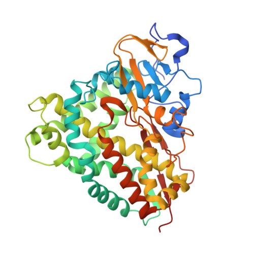
3LXH

Crystal Structure of Cytochrome P450 CYP101D1


Experimental Data Snapshot

  • Method: X-RAY DIFFRACTION
  • Resolution: 2.20 Å
  • R-Value Free: 0.240 
  • R-Value Work: 0.200 
  • R-Value Observed: 0.202 

Starting Model: experimental
View more details

wwPDB Validation   3D Report Full Report


Ligand Structure Quality Assessment 


This is version 1.3 of the entry. See complete history


Literature

Molecular characterization of a class I P450 electron transfer system from Novosphingobium aromaticivorans DSM12444

Yang, W.Bell, S.G.Wang, H.Zhou, W.Hoskins, N.Dale, A.Bartlam, M.Wong, L.L.Rao, Z.

(2010) J Biol Chem 285: 27372-27384

  • DOI: https://doi.org/10.1074/jbc.M110.118349
  • Primary Citation of Related Structures:  
    3LXD, 3LXF, 3LXH, 3LXI

  • PubMed Abstract: 

    Cytochrome P450 (CYP) enzymes of the CYP101 and CYP111 families from the oligotrophic bacterium Novosphingobium aromaticivorans DSM12444 are heme monooxygenases that receive electrons from NADH via Arx, a [2Fe-2S] ferredoxin, and ArR, a ferredoxin reductase. These systems show fast NADH turnovers (k(cat) = 39-91 s(-1)) that are efficiently coupled to product formation. The three-dimensional structures of ArR, Arx, and CYP101D1, which form a physiological class I P450 electron transfer chain, have been resolved by x-ray crystallography. The general structural features of these proteins are similar to their counterparts in other class I systems such as putidaredoxin reductase (PdR), putidaredoxin (Pdx), and CYP101A1 of the camphor hydroxylase system from Pseudomonas putida, and adrenodoxin (Adx) of the mitochondrial steroidogenic CYP11 and CYP24A1 systems. However, significant differences in the proposed protein-protein interaction surfaces of the ferredoxin reductase, ferredoxin, and P450 enzyme are found. There are regions of positive charge on the likely interaction face of ArR and CYP101D1 and a corresponding negatively charged area on the surface of Arx. The [2Fe-2S] cluster binding loop in Arx also has a neutral, hydrophobic patch on the surface. These surface characteristics are more in common with those of Adx than Pdx. The observed structural features are consistent with the ionic strength dependence of the activity.


  • Organizational Affiliation

    Tianjin Key Laboratory of Protein Science, College of Life Sciences, Nankai University, Tianjin 300071, China.


Macromolecules
Find similar proteins by:  (by identity cutoff)  |  3D Structure
Entity ID: 1
MoleculeChains Sequence LengthOrganismDetailsImage
Cytochrome P450
A, B
421Novosphingobium aromaticivorans DSM 12444Mutation(s): 0 
Gene Names: Saro_0514
UniProt
Find proteins for Q2GB12 (Novosphingobium aromaticivorans (strain ATCC 700278 / DSM 12444 / CCUG 56034 / CIP 105152 / NBRC 16084 / F199))
Explore Q2GB12 
Go to UniProtKB:  Q2GB12
Entity Groups  
Sequence Clusters30% Identity50% Identity70% Identity90% Identity95% Identity100% Identity
UniProt GroupQ2GB12
Sequence Annotations
Expand
  • Reference Sequence
Small Molecules
Ligands 3 Unique
IDChains Name / Formula / InChI Key2D Diagram3D Interactions
HEM
Query on HEM

Download Ideal Coordinates CCD File 
C [auth A],
I [auth B]
PROTOPORPHYRIN IX CONTAINING FE
C34 H32 Fe N4 O4
KABFMIBPWCXCRK-RGGAHWMASA-L
PO4
Query on PO4

Download Ideal Coordinates CCD File 
E [auth A]
F [auth A]
G [auth A]
H [auth A]
J [auth B]
E [auth A],
F [auth A],
G [auth A],
H [auth A],
J [auth B],
K [auth B]
PHOSPHATE ION
O4 P
NBIIXXVUZAFLBC-UHFFFAOYSA-K
DIO
Query on DIO

Download Ideal Coordinates CCD File 
D [auth A]1,4-DIETHYLENE DIOXIDE
C4 H8 O2
RYHBNJHYFVUHQT-UHFFFAOYSA-N
Experimental Data & Validation

Experimental Data

  • Method: X-RAY DIFFRACTION
  • Resolution: 2.20 Å
  • R-Value Free: 0.240 
  • R-Value Work: 0.200 
  • R-Value Observed: 0.202 
  • Space Group: P 64 2 2
Unit Cell:
Length ( Å )Angle ( ˚ )
a = 150.902α = 90
b = 150.902β = 90
c = 195.318γ = 120
Software Package:
Software NamePurpose
ADSCdata collection
PHASERphasing
REFMACrefinement
HKL-2000data reduction
HKL-2000data scaling

Structure Validation

View Full Validation Report


Ligand Structure Quality Assessment 


Entry History 

Deposition Data

Revision History  (Full details and data files)

  • Version 1.0: 2010-06-23
    Type: Initial release
  • Version 1.1: 2011-07-13
    Changes: Version format compliance
  • Version 1.2: 2014-02-12
    Changes: Database references
  • Version 1.3: 2023-11-01
    Changes: Data collection, Database references, Derived calculations, Refinement description