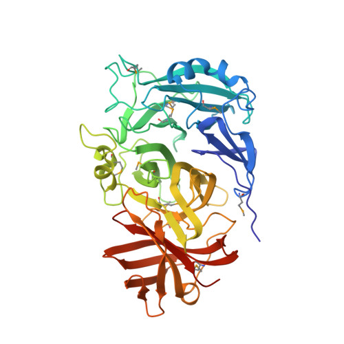
Crystal structure of the glycoside hydrolase, family 43 YxiA protein from Bacillus licheniformis.
Vorobiev, S., Abashidze, M., Seetharaman, J., Belote, R.L., Ciccosanti, C., Sahdev, S., Xiao, R., Acton, T.B., Everett, J.K., Montelione, G.T., Tong, L., Hunt, J.F.To be published.