3LST | pdb_00003lst

Crystal Structure of CalO1, Methyltransferase in Calicheamicin Biosynthesis, SAH bound form


Experimental Data Snapshot

  • Method: X-RAY DIFFRACTION
  • Resolution: 2.40 Å
  • R-Value Free: 
    0.251 (Depositor), 0.247 (DCC) 
  • R-Value Work: 
    0.200 (Depositor), 0.204 (DCC) 
  • R-Value Observed: 
    0.203 (Depositor) 

wwPDB Validation 3D Report Full Report

Validation slider image for 3LST

Ligand Structure Quality Assessment 


This is version 1.3 of the entry. See complete history

Literature

Structural characterization of CalO1: a putative orsellinic acid methyltransferase in the calicheamicin-biosynthetic pathway.

Chang, A.Singh, S.Bingman, C.A.Thorson, J.S.Phillips, G.N.

(2011) Acta Crystallogr D Biol Crystallogr 67: 197-203

  • DOI: https://doi.org/10.1107/S090744491100360X
  • Primary Citation Related Structures: 
    3LST

  • PubMed Abstract: 

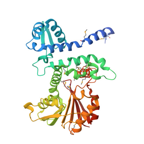
    The X-ray structure determination at 2.4 Å resolution of the putative orsellinic acid C3 O-methyltransferase (CalO1) involved in calicheamicin biosynthesis is reported. Comparison of CalO1 with a homology model of the functionally related calicheamicin orsellinic acid C2 O-methyltransferase (CalO6) implicates several residues that are likely to contribute to the regiospecificity of alkylation. Consistent with the proposed requirement of an acyl-carrier-protein-bound substrate, this structural study also reveals structural determinants within CalO1 that are anticipated to accommodate an association with an acyl carrier protein.


  • Organizational Affiliation
    • Department of Biochemistry, University of Wisconsin-Madison, Madison, Wisconsin 53706, USA.

Macromolecule Content 

  • Total Structure Weight: 76.77 kDa 
  • Atom Count: 5,303 
  • Modeled Residue Count: 666 
  • Deposited Residue Count: 696 
  • Unique protein chains: 1

Macromolecules

Find similar proteins by:|  3D Structure
Entity ID: 1
MoleculeChains  Sequence LengthOrganismDetailsImage
CalO1 Methyltransferase
A, B
348Micromonospora echinosporaMutation(s): 0 
Gene Names: calO1
UniProt
Find proteins for Q8KNE5 (Micromonospora echinospora)
Explore Q8KNE5 
Go to UniProtKB:  Q8KNE5
Entity Groups
Sequence Clusters30% Identity50% Identity70% Identity90% Identity95% Identity100% Identity
UniProt GroupQ8KNE5
Sequence Annotations
Expand
Reference Sequence

Experimental Data & Validation

Experimental Data

  • Method: X-RAY DIFFRACTION
  • Resolution: 2.40 Å
  • R-Value Free:  0.251 (Depositor), 0.247 (DCC) 
  • R-Value Work:  0.200 (Depositor), 0.204 (DCC) 
  • R-Value Observed: 0.203 (Depositor) 
Space Group: C 2 2 21
Unit Cell:
Length ( Å )Angle ( ˚ )
a = 63.515α = 90
b = 93.603β = 90
c = 240.971γ = 90
Software Package:
Software NamePurpose
DENZOdata reduction
SCALEPACKdata scaling
SHARPphasing
PHENIXrefinement
PDB_EXTRACTdata extraction
HKL-2000data collection
HKL-2000data reduction
HKL-2000data scaling

Structure Validation

View Full Validation Report


Ligand Structure Quality Assessment 


Entry History 

Deposition Data

Revision History  (Full details and data files)

  • Version 1.0: 2010-03-02
    Type: Initial release
  • Version 1.1: 2011-07-13
    Changes: Version format compliance
  • Version 1.2: 2017-11-01
    Changes: Refinement description
  • Version 1.3: 2024-10-16
    Changes: Data collection, Database references, Derived calculations, Structure summary