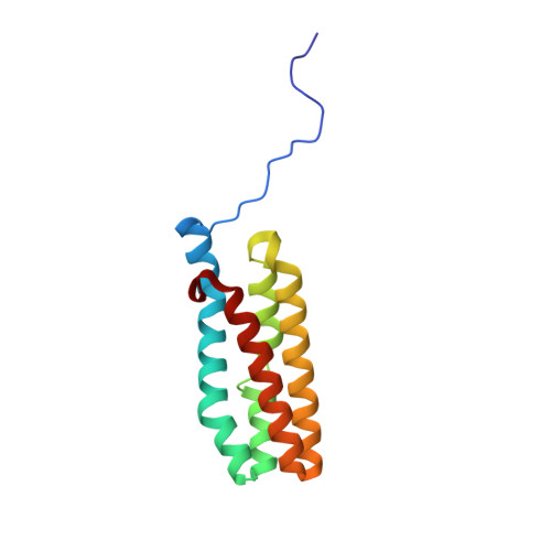
3LS1

Crystal Structure of Cyanobacterial PsbQ from Synechocystis sp. PCC 6803 complexed with Zn2+


Experimental Data Snapshot

  • Method: X-RAY DIFFRACTION
  • Resolution: 1.85 Å
  • R-Value Free: 0.218 
  • R-Value Work: 0.179 
  • R-Value Observed: 0.181 

Starting Model: experimental
View more details

wwPDB Validation   3D Report Full Report


This is version 1.2 of the entry. See complete history


Literature

Crystal Structure of PsbQ from Synechocystis sp. PCC 6803 at 1.8 A: Implications for Binding and Function in Cyanobacterial Photosystem II

Jackson, S.A.Fagerlund, R.D.Wilbanks, S.M.Eaton-Rye, J.J.

(2010) Biochemistry 49: 2765-2767

  • DOI: https://doi.org/10.1021/bi100217h
  • Primary Citation of Related Structures:  
    3LS0, 3LS1

  • PubMed Abstract: 

    In Synechocystis sp. PCC 6803, PsbQ is associated with photosystem II (PSII) complexes with the highest activity and stability. However, this subunit is not found in PSII X-ray crystallographic structures from Thermosynechococcus elongatus or Thermosynechococcus vulcanus. We present the crystal structure of cyanobacterial PsbQ determined in the presence and absence of Zn(2+). The protein has a well-defined helical core, containing four helices arranged in an up-down-up-down fold. A conserved potential interaction site composed of a divalent metal binding site and adjacent hydrophobic pocket has been identified.


  • Organizational Affiliation

    Department of Biochemistry, University of Otago, Dunedin 9054, New Zealand.


Macromolecules
Find similar proteins by:  (by identity cutoff)  |  3D Structure
Entity ID: 1
MoleculeChains Sequence LengthOrganismDetailsImage
Sll1638 protein
A, B
133Synechocystis sp. PCC 6803Mutation(s): 0 
Gene Names: sll1638
UniProt
Find proteins for P73048 (Synechocystis sp. (strain PCC 6803 / Kazusa))
Explore P73048 
Go to UniProtKB:  P73048
Entity Groups  
Sequence Clusters30% Identity50% Identity70% Identity90% Identity95% Identity100% Identity
UniProt GroupP73048
Sequence Annotations
Expand
  • Reference Sequence
Experimental Data & Validation

Experimental Data

  • Method: X-RAY DIFFRACTION
  • Resolution: 1.85 Å
  • R-Value Free: 0.218 
  • R-Value Work: 0.179 
  • R-Value Observed: 0.181 
  • Space Group: I 1 2 1
Unit Cell:
Length ( Å )Angle ( ˚ )
a = 92.771α = 90
b = 29.03β = 97.77
c = 99.503γ = 90
Software Package:
Software NamePurpose
SCALAdata scaling
PHASERphasing
REFMACrefinement
PDB_EXTRACTdata extraction
MOSFLMdata reduction

Structure Validation

View Full Validation Report


Entry History 

Deposition Data

Revision History  (Full details and data files)

  • Version 1.0: 2010-03-31
    Type: Initial release
  • Version 1.1: 2011-07-13
    Changes: Version format compliance
  • Version 1.2: 2023-11-01
    Changes: Data collection, Database references, Derived calculations, Refinement description