3LRP | pdb_00003lrp

Crystal Structure of Plasmodium falciparum ADP-Ribosylation Factor 1


Experimental Data Snapshot

  • Method: X-RAY DIFFRACTION
  • Resolution: 2.50 Å
  • R-Value Free: 
    0.253 (Depositor), 0.250 (DCC) 
  • R-Value Work: 
    0.189 (Depositor), 0.190 (DCC) 
  • R-Value Observed: 
    0.189 (Depositor) 

Starting Model: experimental
View more details

wwPDB Validation   3D Report Full Report


Ligand Structure Quality Assessment 

Created with Raphaël 2.3.0Worse 01 BetterLigand structure goodness of fit to experimental dataBest fitted GDPClick on this verticalbar to view details

This is version 1.3 of the entry. See complete history


Literature

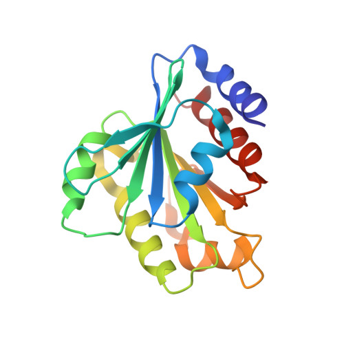
Structure of Plasmodium falciparum ADP-ribosylation factor 1.

Cook, W.J.Smith, C.D.Senkovich, O.Holder, A.A.Chattopadhyay, D.

(2010) Acta Crystallogr Sect F Struct Biol Cryst Commun 66: 1426-1431

  • DOI: https://doi.org/10.1107/S1744309110036997
  • Primary Citation of Related Structures:  
    3LRP

  • PubMed Abstract: 

    Vesicular trafficking may play a crucial role in the pathogenesis and survival of the malaria parasite. ADP-ribosylation factors (ARFs) are among the major components of vesicular trafficking pathways in eukaryotes. The crystal structure of ARF1 GTPase from Plasmodium falciparum has been determined in the GDP-bound conformation at 2.5 Å resolution and is compared with the structures of mammalian ARF1s.


  • Organizational Affiliation

    University of Alabama at Birmingham, Birmingham, AL 35294, USA.


Macromolecules
Find similar proteins by:  (by identity cutoff)  |  3D Structure
Entity ID: 1
MoleculeChains Sequence LengthOrganismDetailsImage
ADP-ribosylation factor 1181Plasmodium falciparumMutation(s): 0 
Gene Names: ARF1ARFPLARF
EC: 3.6.5.2
UniProt
Find proteins for Q7KQL3 (Plasmodium falciparum (isolate 3D7))
Explore Q7KQL3 
Go to UniProtKB:  Q7KQL3
Entity Groups  
Sequence Clusters30% Identity50% Identity70% Identity90% Identity95% Identity100% Identity
UniProt GroupQ7KQL3
Sequence Annotations
Expand
  • Reference Sequence
Experimental Data & Validation

Experimental Data

  • Method: X-RAY DIFFRACTION
  • Resolution: 2.50 Å
  • R-Value Free:  0.253 (Depositor), 0.250 (DCC) 
  • R-Value Work:  0.189 (Depositor), 0.190 (DCC) 
  • R-Value Observed: 0.189 (Depositor) 
Space Group: C 2 2 21
Unit Cell:
Length ( Å )Angle ( ˚ )
a = 45.732α = 90
b = 147.28β = 90
c = 64.229γ = 90
Software Package:
Software NamePurpose
HKL-2000data collection
AMoREphasing
CNSrefinement
HKL-2000data reduction
SCALEPACKdata scaling

Structure Validation

View Full Validation Report


Ligand Structure Quality Assessment 

Created with Raphaël 2.3.0Worse 01 BetterLigand structure goodness of fit to experimental dataBest fitted GDPClick on this verticalbar to view details

Entry History 

Deposition Data

Revision History  (Full details and data files)

  • Version 1.0: 2010-11-10
    Type: Initial release
  • Version 1.1: 2011-07-13
    Changes: Version format compliance
  • Version 1.2: 2013-06-19
    Changes: Database references
  • Version 1.3: 2023-09-06
    Changes: Data collection, Database references, Derived calculations, Refinement description