3LQA | pdb_00003lqa

Crystal structure of clade C gp120 in complex with sCD4 and 21c Fab


Experimental Data Snapshot

  • Method: X-RAY DIFFRACTION
  • Resolution: 3.40 Å
  • R-Value Free: 
    0.322 (Depositor), 0.257 (DCC) 
  • R-Value Work: 
    0.234 (Depositor), 0.241 (DCC) 

wwPDB Validation 3D Report Full Report

Validation slider image for 3LQA

Ligand Structure Quality Assessment 


This is version 1.6 of the entry. See complete history

Literature

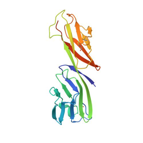
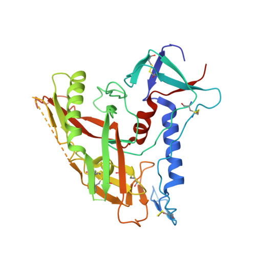
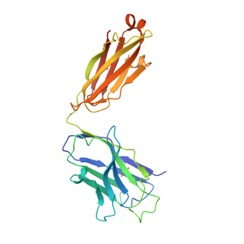
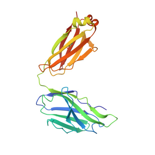
Structure of a clade C HIV-1 gp120 bound to CD4 and CD4-induced antibody reveals anti-CD4 polyreactivity.

Diskin, R.Marcovecchio, P.M.Bjorkman, P.J.

(2010) Nat Struct Mol Biol 17: 608-613

  • DOI: https://doi.org/10.1038/nsmb.1796
  • Primary Citation Related Structures: 
    3LMJ, 3LQA

  • PubMed Abstract: 

    Strategies to combat HIV-1 require structural knowledge of envelope proteins from viruses in HIV-1 clade C, the most rapidly spreading subtype in the world. We present a crystal structure containing a clade C gp120 envelope. The structure, a complex between gp120, the host receptor CD4 and the CD4-induced antibody 21c, reveals that the 21c epitope involves contacts with gp120, a nonself antigen, and with CD4, an autoantigen. Binding studies using wild-type and mutant CD4 show that 21c Fab binds CD4 in the absence of gp120, and that binding of 21c to clade C and HIV-2 gp120s requires the crystallographically observed 21c-CD4 interaction. Additional binding data suggest a role for the gp120 V1V2 loop in creating a high-affinity, but slow-forming, epitope for 21c after CD4 binds. These results contribute to a molecular understanding of CD4-induced antibodies and provide the first visualization to our knowledge of a potentially autoreactive antibody Fab complexed with both self and nonself antigens.


  • Organizational Affiliation
    • Division of Biology, California Institute of Technology, Pasadena, California, USA.

Macromolecule Content 

  • Total Structure Weight: 106.59 kDa 
  • Atom Count: 6,945 
  • Modeled Residue Count: 903 
  • Deposited Residue Count: 972 
  • Unique protein chains: 4

Macromolecules

Find similar proteins by:|  3D Structure
Entity ID: 1
MoleculeChains  Sequence LengthOrganismDetailsImage
T-cell surface glycoprotein CD4A [auth C]192Homo sapiensMutation(s): 0 
Gene Names: CD4
UniProt & NIH Common Fund Data Resources
Find proteins for P01730 (Homo sapiens)
Explore P01730 
Go to UniProtKB:  P01730
PHAROS:  P01730
GTEx:  ENSG00000010610 
Entity Groups
Sequence Clusters30% Identity50% Identity70% Identity90% Identity95% Identity100% Identity
UniProt GroupP01730
Sequence Annotations
Expand
Reference Sequence
Find similar proteins by:|  3D Structure
Entity ID: 2
MoleculeChains  Sequence LengthOrganismDetailsImage
Envelope glycoprotein gp160B [auth G]332Human immunodeficiency virus 1Mutation(s): 7 
Gene Names: env
UniProt
Find proteins for Q1PHM6 (Human immunodeficiency virus type 1)
Explore Q1PHM6 
Go to UniProtKB:  Q1PHM6
Entity Groups
Sequence Clusters30% Identity50% Identity70% Identity90% Identity95% Identity100% Identity
UniProt GroupQ1PHM6
Glycosylation
Glycosylation Sites: 4
Sequence Annotations
Expand
Reference Sequence
Find similar proteins by:|  3D Structure
Entity ID: 3
MoleculeChains  Sequence LengthOrganismDetailsImage
Heavy chain of anti HIV Fab from human 21c antibodyC [auth H]231Homo sapiensMutation(s): 0 
Find similar proteins by:|  3D Structure
Entity ID: 4
MoleculeChains  Sequence LengthOrganismDetailsImage
Light chain of anti HIV Fab from human 21c antibodyD [auth L]217Homo sapiensMutation(s): 0 

Experimental Data & Validation

Experimental Data

  • Method: X-RAY DIFFRACTION
  • Resolution: 3.40 Å
  • R-Value Free:  0.322 (Depositor), 0.257 (DCC) 
  • R-Value Work:  0.234 (Depositor), 0.241 (DCC) 
Space Group: C 2 2 21
Unit Cell:
Length ( Å )Angle ( ˚ )
a = 93.73α = 90
b = 187.93β = 90
c = 151.67γ = 90
Software Package:
Software NamePurpose
CNSrefinement
PDB_EXTRACTdata extraction
Blu-Icedata collection
MOSFLMdata reduction
SCALAdata scaling
PHASERphasing

Structure Validation

View Full Validation Report


Ligand Structure Quality Assessment 


Entry History 

Deposition Data

Revision History  (Full details and data files)

  • Version 1.0: 2010-04-07
    Type: Initial release
  • Version 1.1: 2011-07-13
    Changes: Version format compliance
  • Version 1.2: 2017-08-23
    Changes: Source and taxonomy
  • Version 1.3: 2017-11-01
    Changes: Refinement description
  • Version 1.4: 2020-07-29
    Type: Remediation
    Reason: Carbohydrate remediation
    Changes: Advisory, Data collection, Database references, Derived calculations, Structure summary
  • Version 1.5: 2021-10-13
    Changes: Database references, Structure summary
  • Version 1.6: 2024-10-30
    Changes: Data collection, Structure summary