


Crystal structure of substrate-binding periplasmic protein (Pbp) from Ralstonia solanacearum
Palani, K., Burley, S.K., Swaminathan, S.To be published.
Experimental Data Snapshot
wwPDB Validation 3D Report Full Report

Entity ID: 1 | |||||
|---|---|---|---|---|---|
| Molecule | Chains | Sequence Length | Organism | Details | Image |
| Substrate binding periplasmic protein | 364 | Ralstonia pseudosolanacearum GMI1000 | Mutation(s): 0 Gene Names: RSIPO_02814 | 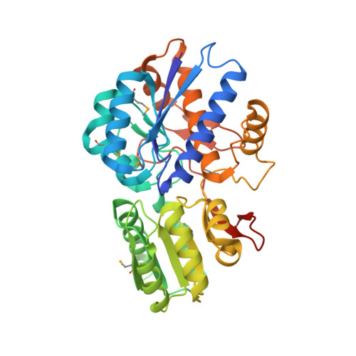 | |
UniProt | |||||
Entity Groups | |||||
| Sequence Clusters | 30% Identity50% Identity70% Identity90% Identity95% Identity100% Identity | ||||
| UniProt Group | Q8XUX2 | ||||
Sequence AnnotationsExpand | |||||
Reference Sequence | |||||
| Ligands 5 Unique | |||||
|---|---|---|---|---|---|
| ID | Chains | Name / Formula / InChI Key | 2D Diagram | 3D Interactions | |
| LEU Download:Ideal Coordinates CCD File | B [auth A] | LEUCINE C6 H13 N O2 ROHFNLRQFUQHCH-YFKPBYRVSA-N |  | ||
| EDO Download:Ideal Coordinates CCD File | C [auth A], D [auth A], E [auth A], F [auth A], G [auth A] | 1,2-ETHANEDIOL C2 H6 O2 LYCAIKOWRPUZTN-UHFFFAOYSA-N |  | ||
| NI Download:Ideal Coordinates CCD File | H [auth A], I [auth A] | NICKEL (II) ION Ni VEQPNABPJHWNSG-UHFFFAOYSA-N |  | ||
| MN Download:Ideal Coordinates CCD File | K [auth A] | MANGANESE (II) ION Mn WAEMQWOKJMHJLA-UHFFFAOYSA-N |  | ||
| MG Download:Ideal Coordinates CCD File | J [auth A] | MAGNESIUM ION Mg JLVVSXFLKOJNIY-UHFFFAOYSA-N |  | ||
| Modified Residues 1 Unique | |||||
|---|---|---|---|---|---|
| ID | Chains | Type | Formula | 2D Diagram | Parent |
| MSE Query on MSE | A | L-PEPTIDE LINKING | C5 H11 N O2 Se |  | MET |
| Length ( Å ) | Angle ( ˚ ) |
|---|---|
| a = 44.11 | α = 90 |
| b = 64.739 | β = 90 |
| c = 114.085 | γ = 90 |
| Software Name | Purpose |
|---|---|
| CNS | refinement |
| CBASS | data collection |
| HKL-2000 | data reduction |
| HKL-2000 | data scaling |
| SHELXD | phasing |
| SHARP | phasing |
| ARP/wARP | model building |


