3LO5 | pdb_00003lo5

Crystal Structure of the dominant negative S17N mutant of Ras


Experimental Data Snapshot

  • Method: X-RAY DIFFRACTION
  • Resolution: 2.57 Å
  • R-Value Free: 
    0.296 (Depositor), 0.294 (DCC) 
  • R-Value Work: 
    0.238 (Depositor), 0.239 (DCC) 
  • R-Value Observed: 
    0.241 (Depositor) 

Starting Model: experimental
View more details

wwPDB Validation 3D Report Full Report

Validation slider image for 3LO5

Ligand Structure Quality Assessment 


This is version 1.3 of the entry. See complete history

Literature

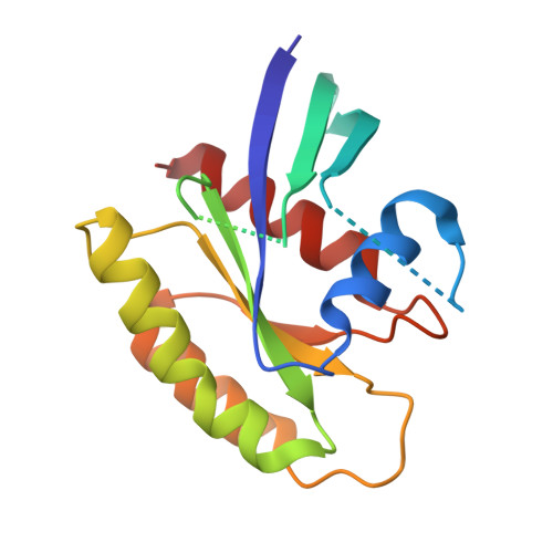
Structure of the Dominant Negative S17N Mutant of Ras

Nassar, N.Singh, K.Garcia-Diaz, M.

(2010) Biochemistry 49: 1970-1974

  • DOI: https://doi.org/10.1021/bi9020742
  • Primary Citation Related Structures: 
    3LO5

  • PubMed Abstract: 

    The use of the dominant negative mutant of Ras has been crucial in elucidating the cellular signaling of Ras in response to the activation of various membrane-bound receptors. Although several point mutants of Ras exhibit a dominant negative effect, the asparagine to serine mutation at position 17 (S17N) remains the most popular and the most effective at inhibiting the activation of endogenous Ras. It is now widely accepted that the dominant negative effect is due to the ability of the mutant to sequester upstream activators and its inability to activate downstream effectors. Here, we present the crystal structure of RasS17N in the GDP-bound form. In the three molecules that populate the asymmetric unit, the Mg(2+) ion that normally coordinates the beta-phosphate is absent because of steric hindrance from the Asn17 side chain. Instead, a Ca(2+) ion is coordinating the alpha-phosphate. Also absent from one molecule is electron density for Phe28, a conserved residue that normally stabilizes the nucleotide's guanine base. Except for Phe28, the nucleotide makes conserved interactions with Ras. Combined, the inability of Phe28 to stabilize the guanine base and the absence of a Mg(2+) ion to neutralize the negative charges on the phosphates explain the weaker affinity of GDP for Ras. Our data suggest that the absence of the Mg(2+) should also dramatically affect GTP binding to Ras and the proper positioning of Thr35 necessary for the activation of switch 1 and the binding to downstream effectors, a prerequisite for the triggering of signaling pathways.


  • Organizational Affiliation
    • Department of Physiology and Biophysics, Stony Brook University, Stony Brook, New York 11794-8661, USA. nicolas.nassar@sunysb.edu

Macromolecule Content 

  • Total Structure Weight: 58.29 kDa 
  • Atom Count: 3,658 
  • Modeled Residue Count: 450 
  • Deposited Residue Count: 498 
  • Unique protein chains: 1

Macromolecules

Find similar proteins by:|  3D Structure
Entity ID: 1
MoleculeChains  Sequence LengthOrganismDetailsImage
GTPase HRasA,
B [auth C],
C [auth E]
166Homo sapiensMutation(s): 1 
Gene Names: H-RasHRASHRAS1
EC: 3.6.5.2
UniProt & NIH Common Fund Data Resources
Find proteins for P01112 (Homo sapiens)
Explore P01112 
Go to UniProtKB:  P01112
PHAROS:  P01112
GTEx:  ENSG00000174775 
Entity Groups
Sequence Clusters30% Identity50% Identity70% Identity90% Identity95% Identity100% Identity
UniProt GroupP01112
Sequence Annotations
Expand
Reference Sequence

Small Molecules

Ligands 3 Unique
IDChains Name / Formula / InChI Key2D Diagram3D Interactions
GDP

Query on GDP


Download:Ideal Coordinates CCD File
D [auth A],
F [auth C],
I [auth E]
GUANOSINE-5'-DIPHOSPHATE
C10 H15 N5 O11 P2
QGWNDRXFNXRZMB-UUOKFMHZSA-N
SO4

Query on SO4


Download:Ideal Coordinates CCD File
G [auth C]SULFATE ION
O4 S
QAOWNCQODCNURD-UHFFFAOYSA-L
CA

Query on CA


Download:Ideal Coordinates CCD File
E [auth A],
H [auth C],
J [auth E],
K [auth E]
CALCIUM ION
Ca
BHPQYMZQTOCNFJ-UHFFFAOYSA-N

Experimental Data & Validation

Experimental Data

  • Method: X-RAY DIFFRACTION
  • Resolution: 2.57 Å
  • R-Value Free:  0.296 (Depositor), 0.294 (DCC) 
  • R-Value Work:  0.238 (Depositor), 0.239 (DCC) 
  • R-Value Observed: 0.241 (Depositor) 
Space Group: I 2 2 2
Unit Cell:
Length ( Å )Angle ( ˚ )
a = 92.475α = 90
b = 102.184β = 90
c = 117.82γ = 90
Software Package:
Software NamePurpose
ADSCdata collection
MOLREPphasing
PHENIXrefinement
HKL-2000data reduction
HKL-2000data scaling

Structure Validation

View Full Validation Report


Ligand Structure Quality Assessment 


Entry History 

Deposition Data

Revision History  (Full details and data files)

  • Version 1.0: 2010-03-02
    Type: Initial release
  • Version 1.1: 2011-07-13
    Changes: Version format compliance
  • Version 1.2: 2021-10-13
    Changes: Database references, Derived calculations
  • Version 1.3: 2023-09-06
    Changes: Data collection, Refinement description