3L6C

X-ray crystal structure of rat serine racemase in complex with malonate a potent inhibitor


Experimental Data Snapshot

  • Method: X-RAY DIFFRACTION
  • Resolution: 2.20 Å
  • R-Value Free: 0.267 
  • R-Value Work: 0.216 
  • R-Value Observed: 0.219 

Starting Model: experimental
View more details

wwPDB Validation   3D Report Full Report


Ligand Structure Quality Assessment 


This is version 1.3 of the entry. See complete history


Literature

The structure of mammalian serine racemase: evidence for conformational changes upon inhibitor binding.

Smith, M.A.Mack, V.Ebneth, A.Moraes, I.Felicetti, B.Wood, M.Schonfeld, D.Mather, O.Cesura, A.Barker, J.

(2010) J Biol Chem 285: 12873-12881

  • DOI: https://doi.org/10.1074/jbc.M109.050062
  • Primary Citation of Related Structures:  
    3HMK, 3L6B, 3L6C, 3L6R

  • PubMed Abstract: 

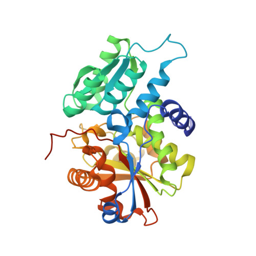
    Serine racemase is responsible for the synthesis of D-serine, an endogenous co-agonist for N-methyl-D-aspartate receptor-type glutamate receptors (NMDARs). This pyridoxal 5'-phosphate-dependent enzyme is involved both in the reversible conversion of L- to D-serine and serine catabolism by alpha,beta-elimination of water, thereby regulating D-serine levels. Because D-serine affects NMDAR signaling throughout the brain, serine racemase is a promising target for the treatment of disorders related to NMDAR dysfunction. To provide a molecular basis for rational drug design the x-ray crystal structures of human and rat serine racemase were determined at 1.5- and 2.1-A resolution, respectively, and in the presence and absence of the orthosteric inhibitor malonate. The structures revealed a fold typical of beta-family pyridoxal 5'-phosphate enzymes, with both a large domain and a flexible small domain associated into a symmetric dimer, and indicated a ligand-induced rearrangement of the small domain that organizes the active site for specific turnover of the substrate.


  • Organizational Affiliation

    Department of Structural Biology and Biology, Evotec, 114 Milton Park, Abingdon, Oxon OX14 4SA, United Kingdom. myron.smith@evotec.com


Macromolecules
Find similar proteins by:  (by identity cutoff)  |  3D Structure
Entity ID: 1
MoleculeChains Sequence LengthOrganismDetailsImage
Serine racemase
A, B
339Rattus norvegicusMutation(s): 2 
Gene Names: Srr
EC: 5.1.1.18
UniProt
Find proteins for Q76EQ0 (Rattus norvegicus)
Explore Q76EQ0 
Go to UniProtKB:  Q76EQ0
Entity Groups  
Sequence Clusters30% Identity50% Identity70% Identity90% Identity95% Identity100% Identity
UniProt GroupQ76EQ0
Sequence Annotations
Expand
  • Reference Sequence
Experimental Data & Validation

Experimental Data

  • Method: X-RAY DIFFRACTION
  • Resolution: 2.20 Å
  • R-Value Free: 0.267 
  • R-Value Work: 0.216 
  • R-Value Observed: 0.219 
  • Space Group: P 3
Unit Cell:
Length ( Å )Angle ( ˚ )
a = 110.593α = 90
b = 110.593β = 90
c = 47.917γ = 120
Software Package:
Software NamePurpose
CrystalCleardata collection
PHASERphasing
REFMACrefinement
d*TREKdata reduction
d*TREKdata scaling

Structure Validation

View Full Validation Report


Ligand Structure Quality Assessment 


Entry History 

Deposition Data

Revision History  (Full details and data files)

  • Version 1.0: 2010-01-26
    Type: Initial release
  • Version 1.1: 2011-07-13
    Changes: Version format compliance
  • Version 1.2: 2021-11-10
    Changes: Database references, Derived calculations
  • Version 1.3: 2024-04-03
    Changes: Data collection, Refinement description