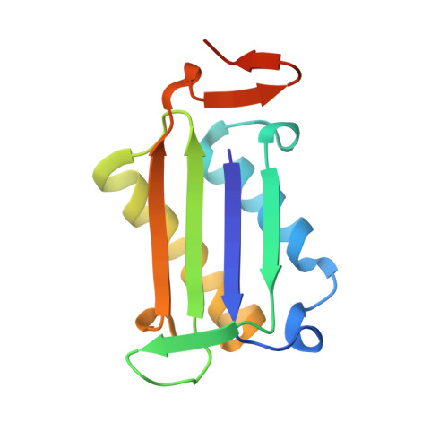
3L5P

Crystal structure of macrophage migration inhibitory factor (MIF) with imidazopyridazinol inhibitor at 1.80A resolution


Experimental Data Snapshot

  • Method: X-RAY DIFFRACTION
  • Resolution: 1.80 Å
  • R-Value Free: 0.221 
  • R-Value Work: 0.198 
  • R-Value Observed: 0.199 

Starting Model: experimental
View more details

wwPDB Validation   3D Report Full Report


Ligand Structure Quality Assessment 


This is version 1.3 of the entry. See complete history


Literature

Fragment screening of inhibitors for MIF tautomerase reveals a cryptic surface binding site.

McLean, L.R.Zhang, Y.Li, H.Choi, Y.M.Han, Z.Vaz, R.J.Li, Y.

(2010) Bioorg Med Chem Lett 20: 1821-1824

  • DOI: https://doi.org/10.1016/j.bmcl.2010.02.009
  • Primary Citation of Related Structures:  
    3L5P, 3L5R, 3L5S, 3L5T, 3L5U, 3L5V

  • PubMed Abstract: 

    In the course of a fragment screening campaign by in silico docking followed by X-ray crystallography, a novel binding site for migration inhibitory factor (MIF) inhibitors was demonstrated. The site is formed by rotation of the side-chain of Tyr-36 to reveal a surface binding site in MIF that is hydrophobic and surrounded by aromatic side-chain residues. The crystal structures of two small inhibitors that bind to this site and of a quinolinone inhibitor, that spans the canonical deep pocket near Pro-1 and the new surface binding site, have been solved. These results suggest new opportunities for structure-based design of MIF inhibitors.


  • Organizational Affiliation

    Discovery Research, Sanofi-aventis, Bridgewater, NJ 08807, USA. Larry.Mclean@sanofi-aventis.com


Macromolecules
Find similar proteins by:  (by identity cutoff)  |  3D Structure
Entity ID: 1
MoleculeChains Sequence LengthOrganismDetailsImage
Macrophage migration inhibitory factor
A, B, C
122Homo sapiensMutation(s): 0 
Gene Names: GLIFMIFMMIF
EC: 5.3.2.1 (PDB Primary Data), 5.3.3.12 (PDB Primary Data)
UniProt & NIH Common Fund Data Resources
Find proteins for P14174 (Homo sapiens)
Explore P14174 
Go to UniProtKB:  P14174
GTEx:  ENSG00000240972 
Entity Groups  
Sequence Clusters30% Identity50% Identity70% Identity90% Identity95% Identity100% Identity
UniProt GroupP14174
Sequence Annotations
Expand
  • Reference Sequence
Experimental Data & Validation

Experimental Data

  • Method: X-RAY DIFFRACTION
  • Resolution: 1.80 Å
  • R-Value Free: 0.221 
  • R-Value Work: 0.198 
  • R-Value Observed: 0.199 
  • Space Group: P 21 21 21
Unit Cell:
Length ( Å )Angle ( ˚ )
a = 67.675α = 90
b = 67.854β = 90
c = 89.009γ = 90
Software Package:
Software NamePurpose
DENZOdata reduction
SCALEPACKdata scaling
MOLREPphasing
CNSrefinement
PDB_EXTRACTdata extraction
CrystalCleardata collection
CNXrefinement

Structure Validation

View Full Validation Report


Ligand Structure Quality Assessment 


Entry History 

Deposition Data

Revision History  (Full details and data files)

  • Version 1.0: 2010-03-09
    Type: Initial release
  • Version 1.1: 2011-07-13
    Changes: Version format compliance
  • Version 1.2: 2017-11-01
    Changes: Advisory, Refinement description
  • Version 1.3: 2023-09-06
    Changes: Advisory, Data collection, Database references, Derived calculations, Refinement description