3L3N | pdb_00003l3n

Testis ACE co-crystal structure with novel inhibitor lisW


Experimental Data Snapshot

  • Method: X-RAY DIFFRACTION
  • Resolution: 2.30 Å
  • R-Value Free: 
    0.266 (Depositor), 0.267 (DCC) 
  • R-Value Work: 
    0.226 (Depositor), 0.229 (DCC) 

Starting Model: experimental
View more details

wwPDB Validation 3D Report Full Report

Validation slider image for 3L3N

Ligand Structure Quality Assessment 


This is version 2.3 of the entry. See complete history

Literature

Characterization of domain-selective inhibitor binding in angiotensin-converting enzyme using a novel derivative of lisinopril.

Watermeyer, J.M.Kroger, W.L.O'Neill, H.G.Sewell, B.T.Sturrock, E.D.

(2010) Biochem J 428: 67-74

  • DOI: https://doi.org/10.1042/BJ20100056
  • Primary Citation Related Structures: 
    3L3N

  • PubMed Abstract: 

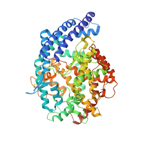
    Human ACE (angiotensin-converting enzyme) (EC 3.4.15.1) is an important drug target because of its role in the regulation of blood pressure via the renin-angiotensin-aldosterone system. Somatic ACE comprises two homologous domains, the differing substrate preferences of which present a new avenue for domain-selective inhibitor design. We have co-crystallized lisW-S, a C-domain-selective derivative of the drug lisinopril, with human testis ACE and determined a structure using X-ray crystallography to a resolution of 2.30 A (1 A=0.1 nm). In this structure, lisW-S is seen to have a similar binding mode to its parent compound lisinopril, but the P2' tryptophan moiety takes a different conformation to that seen in other inhibitors having a tryptophan residue in this position. We have examined further the domain-specific interactions of this inhibitor by mutating C-domain-specific active-site residues to their N domain equivalents, then assessing the effect of the mutation on inhibition by lisW-S using a fluorescence-based assay. Kinetics analysis shows a 258-fold domain-selectivity that is largely due to the co-operative effect of C-domain-specific residues in the S2' subsite. The high affinity and selectivity of this inhibitor make it a good lead candidate for cardiovascular drug development.


  • Organizational Affiliation
    • Division of Medical Biochemistry, Institute of Infectious Disease and Molecular Medicine, University of Cape Town, Observatory, Cape Town, 7935, South Africa.

Macromolecule Content 

  • Total Structure Weight: 69.48 kDa 
  • Atom Count: 4,942 
  • Modeled Residue Count: 575 
  • Deposited Residue Count: 591 
  • Unique protein chains: 1

Macromolecules

Find similar proteins by:|  3D Structure
Entity ID: 1
MoleculeChains  Sequence LengthOrganismDetailsImage
Angiotensin-converting enzyme591Homo sapiensMutation(s): 5 
Gene Names: ACEDCPDCP1
EC: 3.4.15.1 (PDB Primary Data), 3.2.1 (PDB Primary Data)
UniProt & NIH Common Fund Data Resources
Find proteins for P12821 (Homo sapiens)
Explore P12821 
Go to UniProtKB:  P12821
PHAROS:  P12821
GTEx:  ENSG00000159640 
Entity Groups
Sequence Clusters30% Identity50% Identity70% Identity90% Identity95% Identity100% Identity
UniProt GroupP12821
Glycosylation
Glycosylation Sites: 1Go to GlyGen: P12821-1
Sequence Annotations
Expand
Reference Sequence

Oligosaccharides

Help  
Entity ID: 2
MoleculeChains Length2D Diagram GlycosylationD Interactions
2-acetamido-2-deoxy-beta-D-glucopyranose-(1-4)-[alpha-L-fucopyranose-(1-6)]2-acetamido-2-deoxy-beta-D-glucopyranose
B
3N-Glycosylation
Glycosylation Resources
GlyTouCan: G21290RB
GlyCosmos: G21290RB
GlyGen: G21290RB

Small Molecules

Experimental Data & Validation

Experimental Data

  • Method: X-RAY DIFFRACTION
  • Resolution: 2.30 Å
  • R-Value Free:  0.266 (Depositor), 0.267 (DCC) 
  • R-Value Work:  0.226 (Depositor), 0.229 (DCC) 
Space Group: P 21 21 21
Unit Cell:
Length ( Å )Angle ( ˚ )
a = 56.383α = 90
b = 84.789β = 90
c = 133.958γ = 90
Software Package:
Software NamePurpose
DENZOdata reduction
SCALEPACKdata scaling
EPMRphasing
CNSrefinement
PDB_EXTRACTdata extraction
HKL-2000data collection
HKL-2000data reduction

Structure Validation

View Full Validation Report


Ligand Structure Quality Assessment 


Entry History 

Deposition Data

Revision History  (Full details and data files)

  • Version 1.0: 2010-04-28
    Type: Initial release
  • Version 1.1: 2011-07-13
    Changes: Version format compliance
  • Version 1.2: 2017-11-01
    Changes: Refinement description
  • Version 2.0: 2020-07-29
    Type: Remediation
    Reason: Carbohydrate remediation
    Changes: Atomic model, Data collection, Derived calculations, Structure summary
  • Version 2.1: 2021-10-13
    Changes: Database references, Structure summary
  • Version 2.2: 2023-09-06
    Changes: Data collection, Refinement description
  • Version 2.3: 2024-10-30
    Changes: Structure summary