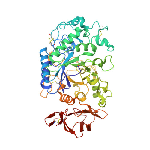
3L2M

X-ray Crystallographic Analysis of Pig Pancreatic Alpha-Amylase with Alpha-cyclodextrin


Experimental Data Snapshot

  • Method: X-RAY DIFFRACTION
  • Resolution: 1.97 Å
  • R-Value Free: 0.160 
  • R-Value Work: 0.127 
  • R-Value Observed: 0.130 

wwPDB Validation   3D Report Full Report


This is version 3.1 of the entry. See complete history


Literature

X-ray crystallographic analyses of pig pancreatic alpha-amylase with limit dextrin, oligosaccharide, and alpha-cyclodextrin.

Larson, S.B.Day, J.S.McPherson, A.

(2010) Biochemistry 49: 3101-3115

  • DOI: https://doi.org/10.1021/bi902183w
  • Primary Citation of Related Structures:  
    3L2L, 3L2M

  • PubMed Abstract: 

    Further refinement of the model using maximum likelihood procedures and reevaluation of the native electron density map has shown that crystals of pig pancreatic alpha-amylase, whose structure we reported more than 15 years ago, in fact contain a substantial amount of carbohydrate. The carbohydrate fragments are the products of glycogen digestion carried out as an essential step of the protein's purification procedure. In particular, the substrate-binding cleft contains a limit dextrin of six glucose residues, one of which contains both alpha-(1,4) and alpha-(1,6) linkages to contiguous residues. The disaccharide in the original model, shared between two amylase molecules in the crystal lattice, but also occupying a portion of the substrate-binding cleft, is now seen to be a tetrasaccharide. There are, in addition, several other probable monosaccharide binding sites. Furthermore, we have further reviewed our X-ray diffraction analysis of alpha-amylase complexed with alpha-cyclodextrin. alpha-Amylase binds three cyclodextrin molecules. Glucose residues of two of the rings superimpose upon the limit dextrin and the tetrasaccharide. The limit dextrin superimposes in large part upon linear oligosaccharide inhibitors visualized by other investigators. By comprehensive integration of these complexes we have constructed a model for the binding of polysaccharides having the helical character known to be present in natural substrates such as starch and glycogen.


  • Organizational Affiliation

    Department of Molecular Biology and Biochemistry, The University of California, Irvine, California 92697-3900, USA.


Macromolecules
Find similar proteins by:  (by identity cutoff)  |  3D Structure
Entity ID: 1
MoleculeChains Sequence LengthOrganismDetailsImage
Pancreatic alpha-amylase496Sus scrofaMutation(s): 0 
EC: 3.2.1.1
UniProt
Find proteins for P00690 (Sus scrofa)
Explore P00690 
Go to UniProtKB:  P00690
Entity Groups  
Sequence Clusters30% Identity50% Identity70% Identity90% Identity95% Identity100% Identity
UniProt GroupP00690
Sequence Annotations
Expand
  • Reference Sequence
Oligosaccharides

Help

Entity ID: 2
MoleculeChains Length2D Diagram Glycosylation3D Interactions
Cyclohexakis-(1-4)-(alpha-D-glucopyranose)
B, C, D
6N/A
Glycosylation Resources
GlyTouCan:  G22307HL
GlyCosmos:  G22307HL
Small Molecules
Modified Residues  1 Unique
IDChains TypeFormula2D DiagramParent
PCA
Query on PCA
A
L-PEPTIDE LINKINGC5 H7 N O3GLN
Biologically Interesting Molecules (External Reference) 1 Unique
Experimental Data & Validation

Experimental Data

  • Method: X-RAY DIFFRACTION
  • Resolution: 1.97 Å
  • R-Value Free: 0.160 
  • R-Value Work: 0.127 
  • R-Value Observed: 0.130 
  • Space Group: P 21 21 21
Unit Cell:
Length ( Å )Angle ( ˚ )
a = 70.65α = 90
b = 114.88β = 90
c = 118.85γ = 90
Software Package:
Software NamePurpose
SDMSdata collection
REFMACrefinement
SDMSdata reduction
SDMSdata scaling

Structure Validation

View Full Validation Report


Entry History 

Deposition Data

Revision History  (Full details and data files)

  • Version 1.0: 2010-04-14
    Type: Initial release
  • Version 1.1: 2011-07-13
    Changes: Advisory, Version format compliance
  • Version 1.2: 2017-11-01
    Changes: Advisory, Refinement description
  • Version 2.0: 2019-12-25
    Changes: Data collection, Database references, Derived calculations, Polymer sequence
  • Version 3.0: 2020-07-29
    Type: Remediation
    Reason: Carbohydrate remediation
    Changes: Advisory, Atomic model, Data collection, Derived calculations, Structure summary
  • Version 3.1: 2024-10-16
    Changes: Data collection, Database references, Derived calculations, Structure summary