3KXS | pdb_00003kxs

Crystal structure of HBV capsid mutant dimer (oxy form), strain adyw


Experimental Data Snapshot

  • Method: X-RAY DIFFRACTION
  • Resolution: 2.25 Å
  • R-Value Free: 
    0.264 (Depositor), 0.260 (DCC) 
  • R-Value Work: 
    0.217 (Depositor), 0.215 (DCC) 
  • R-Value Observed: 
    0.220 (Depositor) 

Starting Model: experimental
View more details

wwPDB Validation 3D Report Full Report

Validation slider image for 3KXS

This is version 1.4 of the entry. See complete history

Literature

Conformational changes in the hepatitis B virus core protein are consistent with a role for allostery in virus assembly

Packianathan, C.Katen, S.P.Dann, C.E.Zlotnick, A.

(2010) J Virol 84: 1607-1615

  • DOI: https://doi.org/10.1128/JVI.02033-09
  • Primary Citation Related Structures: 
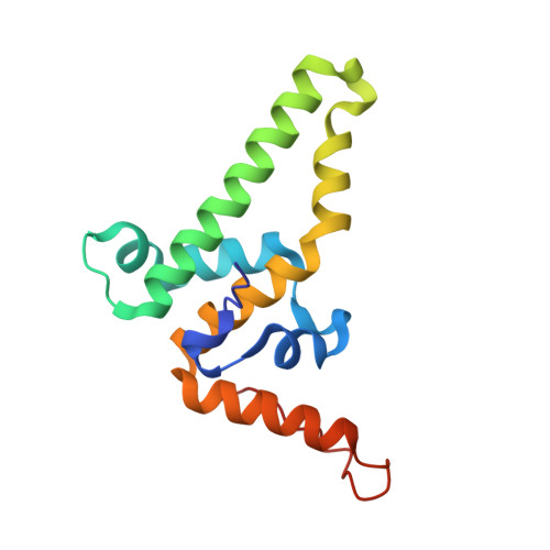
    3KXS

  • PubMed Abstract: 

    In infected cells, virus components must be organized at the right place and time to ensure assembly of infectious virions. From a different perspective, assembly must be prevented until all components are available. Hypothetically, this can be achieved by allosterically controlling assembly. Consistent with this hypothesis, here we show that the structure of the hepatitis B virus (HBV) core protein dimer, which can spontaneously self-assemble, is incompatible with capsid assembly. Systematic differences between core protein dimer and capsid conformations demonstrate linkage between the intradimer interface and interdimer contact surface. These structures also provide explanations for the capsid-dimer selectivity of some antibodies and the activities of assembly effectors. Solution studies suggest that the assembly-inactive state is more accurately an ensemble of conformations. Simulations show that allostery supports controlled assembly and results in capsids that are resistant to dissociation. We propose that allostery, as demonstrated in HBV, is common to most self-assembling viruses.


  • Organizational Affiliation
    • Department of Molecular and Cellular Biochemistry, Indiana University, 212 S. Hawthorne Dr., Bloomington, IN 47405-7003, USA.

Macromolecule Content 

  • Total Structure Weight: 96.39 kDa 
  • Atom Count: 6,932 
  • Modeled Residue Count: 850 
  • Deposited Residue Count: 858 
  • Unique protein chains: 1

Macromolecules

Find similar proteins by:|  3D Structure
Entity ID: 1
MoleculeChains  Sequence LengthOrganismDetailsImage
Capsid protein143Hepatitis B virus subtype adywMutation(s): 1 
Gene Names: C
UniProt
Find proteins for P03147 (Hepatitis B virus genotype D subtype adw (isolate United Kingdom/adyw/1979))
Explore P03147 
Go to UniProtKB:  P03147
Entity Groups
Sequence Clusters30% Identity50% Identity70% Identity90% Identity95% Identity100% Identity
UniProt GroupP03147
Sequence Annotations
Expand
Reference Sequence

Experimental Data & Validation

Experimental Data

  • Method: X-RAY DIFFRACTION
  • Resolution: 2.25 Å
  • R-Value Free:  0.264 (Depositor), 0.260 (DCC) 
  • R-Value Work:  0.217 (Depositor), 0.215 (DCC) 
  • R-Value Observed: 0.220 (Depositor) 
Space Group: P 31
Unit Cell:
Length ( Å )Angle ( ˚ )
a = 104.162α = 90
b = 104.162β = 90
c = 86.917γ = 120
Software Package:
Software NamePurpose
CrystalCleardata collection
PHASERphasing
REFMACrefinement
CNSrefinement
HKL-2000data reduction
HKL-2000data scaling

Structure Validation

View Full Validation Report


Entry History 

Deposition Data

Revision History  (Full details and data files)

  • Version 1.0: 2010-02-02
    Type: Initial release
  • Version 1.1: 2011-07-13
    Changes: Version format compliance
  • Version 1.2: 2021-10-13
    Changes: Database references, Derived calculations
  • Version 1.3: 2023-09-06
    Changes: Data collection, Refinement description
  • Version 1.4: 2024-11-06
    Changes: Structure summary