3KRQ | pdb_00003krq

Crystal structure of the complex of lactoperoxidase with a potent inhibitor amino-triazole at 2.2a resolution


Experimental Data Snapshot

  • Method: X-RAY DIFFRACTION
  • Resolution: 2.25 Å
  • R-Value Free: 
    0.249 (Depositor), 0.264 (DCC) 
  • R-Value Work: 
    0.226 (Depositor), 0.234 (DCC) 
  • R-Value Observed: 
    0.226 (Depositor) 

Starting Model: experimental
View more details

wwPDB Validation 3D Report Full Report

Validation slider image for 3KRQ

Ligand Structure Quality Assessment 


This is version 2.2 of the entry. See complete history

Literature

First structural evidence for the mode of diffusion of aromatic ligands and ligand-induced closure of the hydrophobic channel in heme peroxidases

Singh, A.K.Singh, N.Tiwari, A.Sinha, M.Kushwaha, G.S.Kaur, P.Srinivasan, A.Sharma, S.Singh, T.P.

(2010) J Biol Inorg Chem 15: 1099-1107

  • DOI: https://doi.org/10.1007/s00775-010-0669-3
  • Primary Citation Related Structures: 
    3KRQ

  • PubMed Abstract: 

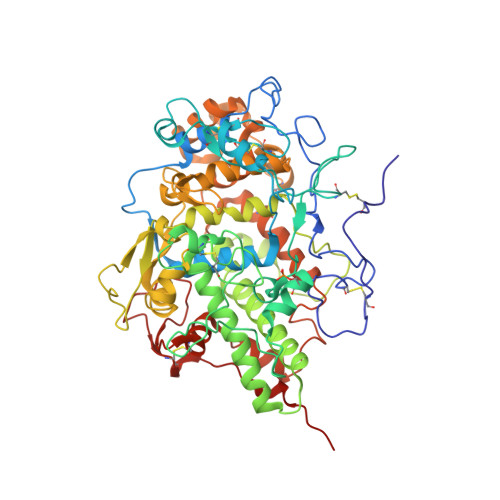
    The mode of binding of aromatic ligands in the substrate binding site on the distal heme side in heme peroxidases is well understood. However, the mode of diffusion through the extended hydrophobic channel and the regulatory role of the channel are not yet clear. To provide answers to these questions, the crystal structure of the complex of lactoperoxidase and 3-amino-1,2,4-triazole (amitrole) has been determined, which revealed the presence of two ligand molecules, one in the substrate binding site and the second in the hydrophobic channel. The binding of ligand in the channel induced a remarkable conformational change in the side chain of Phe254, which flips from its original distant position to interact with the trapped ligand in the hydrophobic channel. As a result, the channel is completely blocked so that no ligand can diffuse through it to the substrate binding site. Another amitrole molecule is bound to lactoperoxidase in the substrate binding site by replacing three water molecules, including the crucial iron-bound water molecule, W1. In this arrangement, the amino nitrogen atom of amitrole occupies the position of W1 and interacts directly with ferric iron. As a consequence, it prevents the binding of H2O2 to heme iron. Thus, the interactions of amitrole with lactoperoxidase obstruct both the passage of ligands through the hydrophobic channel as well as the binding of H2O2. This explains the amitrole toxicity. From binding studies, the dissociation constant (Kd) for amitrole with lactoperoxidase was found to be approximately 5.5x10(-7) M, indicating high affinity.


  • Organizational Affiliation
    • Department of Biophysics, All India Institute of Medical Sciences, Ansari Nagar, New Delhi, 110029, India.

Macromolecule Content 

  • Total Structure Weight: 71.97 kDa 
  • Atom Count: 5,418 
  • Modeled Residue Count: 595 
  • Deposited Residue Count: 595 
  • Unique protein chains: 1

Macromolecules

Find similar proteins by:|  3D Structure
Entity ID: 1
MoleculeChains  Sequence LengthOrganismDetailsImage
Lactoperoxidase595Bos taurusMutation(s): 0 
EC: 1.11.1.7
UniProt
Find proteins for P80025 (Bos taurus)
Explore P80025 
Go to UniProtKB:  P80025
Entity Groups
Sequence Clusters30% Identity50% Identity70% Identity90% Identity95% Identity100% Identity
UniProt GroupP80025
Glycosylation
Glycosylation Sites: 4
Sequence Annotations
Expand
Reference Sequence

Oligosaccharides

Help  
Entity ID: 2
MoleculeChains Length2D Diagram GlycosylationD Interactions
alpha-D-mannopyranose-(1-4)-2-acetamido-2-deoxy-beta-D-glucopyranose-(1-4)-2-acetamido-2-deoxy-beta-D-glucopyranose
B, D
3N-Glycosylation
Glycosylation Resources
GlyTouCan: G62182OO
GlyCosmos: G62182OO
GlyGen: G62182OO
Entity ID: 3
MoleculeChains Length2D Diagram GlycosylationD Interactions
beta-D-mannopyranose-(1-4)-2-acetamido-2-deoxy-beta-D-glucopyranose-(1-4)-2-acetamido-2-deoxy-beta-D-glucopyranose
C
3N-Glycosylation
Glycosylation Resources
GlyTouCan: G15407YE
GlyCosmos: G15407YE
GlyGen: G15407YE
Entity ID: 4
MoleculeChains Length2D Diagram GlycosylationD Interactions
2-acetamido-2-deoxy-alpha-D-glucopyranose-(1-4)-2-acetamido-2-deoxy-beta-D-glucopyranose
E
2N-Glycosylation
Glycosylation Resources
GlyTouCan: G07375KG
GlyCosmos: G07375KG
GlyGen: G07375KG

Small Molecules

Ligands 6 Unique
IDChains Name / Formula / InChI Key2D Diagram3D Interactions
HEM

Query on HEM


Download:Ideal Coordinates CCD File
F [auth A]PROTOPORPHYRIN IX CONTAINING FE
C34 H32 Fe N4 O4
KABFMIBPWCXCRK-RGGAHWMASA-L
IOD

Query on IOD


Download:Ideal Coordinates CCD File
H [auth A]
I [auth A]
J [auth A]
K [auth A]
L [auth A]
H [auth A],
I [auth A],
J [auth A],
K [auth A],
L [auth A],
M [auth A],
N [auth A]
IODIDE ION
I
XMBWDFGMSWQBCA-UHFFFAOYSA-M
MRD

Query on MRD


Download:Ideal Coordinates CCD File
Q [auth A](4R)-2-METHYLPENTANE-2,4-DIOL
C6 H14 O2
SVTBMSDMJJWYQN-RXMQYKEDSA-N
PEG

Query on PEG


Download:Ideal Coordinates CCD File
R [auth A]DI(HYDROXYETHYL)ETHER
C4 H10 O3
MTHSVFCYNBDYFN-UHFFFAOYSA-N
3TR

Query on 3TR


Download:Ideal Coordinates CCD File
O [auth A],
P [auth A]
3-AMINO-1,2,4-TRIAZOLE
C2 H4 N4
KLSJWNVTNUYHDU-UHFFFAOYSA-N
CA

Query on CA


Download:Ideal Coordinates CCD File
G [auth A]CALCIUM ION
Ca
BHPQYMZQTOCNFJ-UHFFFAOYSA-N
Modified Residues  1 Unique
IDChains TypeFormula2D DiagramParent
SEP
Query on SEP
A
L-PEPTIDE LINKINGC3 H8 N O6 PSER

Experimental Data & Validation

Experimental Data

  • Method: X-RAY DIFFRACTION
  • Resolution: 2.25 Å
  • R-Value Free:  0.249 (Depositor), 0.264 (DCC) 
  • R-Value Work:  0.226 (Depositor), 0.234 (DCC) 
  • R-Value Observed: 0.226 (Depositor) 
Space Group: P 1 21 1
Unit Cell:
Length ( Å )Angle ( ˚ )
a = 54.487α = 90
b = 81.105β = 100.82
c = 73.928γ = 90
Software Package:
Software NamePurpose
MAR345dtbdata collection
AMoREphasing
CNSrefinement
AUTOMARdata reduction
SCALEPACKdata scaling

Structure Validation

View Full Validation Report


Ligand Structure Quality Assessment 


Entry History 

Deposition Data

Revision History  (Full details and data files)

  • Version 1.0: 2010-05-26
    Type: Initial release
  • Version 1.1: 2011-07-13
    Changes: Version format compliance
  • Version 1.2: 2011-08-24
    Changes: Database references, Non-polymer description, Structure summary
  • Version 2.0: 2020-07-29
    Type: Remediation
    Reason: Carbohydrate remediation
    Changes: Atomic model, Data collection, Derived calculations, Structure summary
  • Version 2.1: 2023-11-01
    Changes: Data collection, Database references, Refinement description, Structure summary
  • Version 2.2: 2024-11-20
    Changes: Structure summary