NMR derived topology of a GFP-photoprotein energy transfer complex
Titushin, M.S., Feng, Y., Stepanyuk, G.A., Li, Y., Markova, S.V., Golz, S., Wang, B.-C., Lee, J., Wang, J., Vysotski, E.S., Liu, Z.-J.(2010) J Biological Chem 285: 40891-40900
- PubMed: 20926380 Search on PubMedSearch on PubMed Central
- DOI: https://doi.org/10.1074/jbc.M110.133843
- Primary Citation Related Structures:
3KPX - PubMed Abstract:
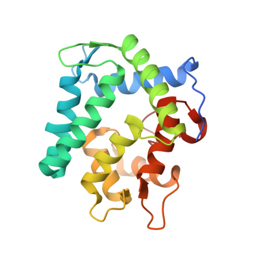
Förster resonance energy transfer within a protein-protein complex has previously been invoked to explain emission spectral modulation observed in several bioluminescence systems. Here we present a spatial structure of a complex of the Ca(2+)-regulated photoprotein clytin with its green-fluorescent protein (cgGFP) from the jellyfish Clytia gregaria, and show that it accounts for the bioluminescence properties of this system in vitro. We adopted an indirect approach of combining x-ray crystallography determined structures of the separate proteins, NMR spectroscopy, computational docking, and mutagenesis. Heteronuclear NMR spectroscopy using variously (15)N,(13)C,(2)H-enriched proteins enabled assignment of backbone resonances of more than 94% of the residues of both proteins. In a mixture of the two proteins at millimolar concentrations, complexation was inferred from perturbations of certain (1)H-(15)N HSQC-resonances, which could be mapped to those residues involved at the interaction site. A docking computation using HADDOCK was employed constrained by the sites of interaction, to deduce an overall spatial structure of the complex. Contacts within the clytin-cgGFP complex and electrostatic complementarity of interaction surfaces argued for a weak protein-protein complex. A weak affinity was also observed by isothermal titration calorimetry (K(D) = 0.9 mM). Mutation of clytin residues located at the interaction site reduced the degree of protein-protein association concomitant with a loss of effectiveness of cgGFP in color-shifting the bioluminescence. It is suggested that this clytin-cgGFP structure corresponds to the transient complex previously postulated to account for the energy transfer effect of GFP in the bioluminescence of aequorin or Renilla luciferase.
- National Laboratory of Biomacromolecules, Institute of Biophysics Chinese Academy of Sciences, Datun Road 15, Beijing 100101, China.
Organizational Affiliation: