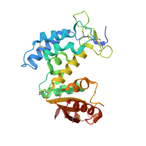
3KOU

Structural insights into the catalytic mechanism of CD38: Evidence for a conformationally flexible covalent enzyme-substrate complex.


Experimental Data Snapshot

  • Method: X-RAY DIFFRACTION
  • Resolution: 1.78 Å
  • R-Value Free: 0.243 
  • R-Value Work: 0.200 
  • R-Value Observed: 0.203 

Starting Model: experimental
View more details

wwPDB Validation   3D Report Full Report


Ligand Structure Quality Assessment 


This is version 2.2 of the entry. See complete history


Literature

Insights into the mechanism of bovine CD38/NAD+glycohydrolase from the X-ray structures of its Michaelis complex and covalently-trapped intermediates.

Egea, P.F.Muller-Steffner, H.Kuhn, I.Cakir-Kiefer, C.Oppenheimer, N.J.Stroud, R.M.Kellenberger, E.Schuber, F.

(2012) PLoS One 7: e34918-e34918

  • DOI: https://doi.org/10.1371/journal.pone.0034918
  • Primary Citation of Related Structures:  
    3GC6, 3GH3, 3GHH, 3KOU, 3P5S

  • PubMed Abstract: 

    Bovine CD38/NAD(+)glycohydrolase (bCD38) catalyses the hydrolysis of NAD(+) into nicotinamide and ADP-ribose and the formation of cyclic ADP-ribose (cADPR). We solved the crystal structures of the mono N-glycosylated forms of the ecto-domain of bCD38 or the catalytic residue mutant Glu218Gln in their apo state or bound to aFNAD or rFNAD, two 2'-fluorinated analogs of NAD(+). Both compounds behave as mechanism-based inhibitors, allowing the trapping of a reaction intermediate covalently linked to Glu218. Compared to the non-covalent (Michaelis) complex, the ligands adopt a more folded conformation in the covalent complexes. Altogether these crystallographic snapshots along the reaction pathway reveal the drastic conformational rearrangements undergone by the ligand during catalysis with the repositioning of its adenine ring from a solvent-exposed position stacked against Trp168 to a more buried position stacked against Trp181. This adenine flipping between conserved tryptophans is a prerequisite for the proper positioning of the N1 of the adenine ring to perform the nucleophilic attack on the C1' of the ribofuranoside ring ultimately yielding cADPR. In all structures, however, the adenine ring adopts the most thermodynamically favorable anti conformation, explaining why cyclization, which requires a syn conformation, remains a rare alternate event in the reactions catalyzed by bCD38 (cADPR represents only 1% of the reaction products). In the Michaelis complex, the substrate is bound in a constrained conformation; the enzyme uses this ground-state destabilization, in addition to a hydrophobic environment and desolvation of the nicotinamide-ribosyl bond, to destabilize the scissile bond leading to the formation of a ribooxocarbenium ion intermediate. The Glu218 side chain stabilizes this reaction intermediate and plays another important role during catalysis by polarizing the 2'-OH of the substrate NAD(+). Based on our structural analysis and data on active site mutants, we propose a detailed analysis of the catalytic mechanism.


  • Organizational Affiliation

    Department of Biological Chemistry, University of California Los Angeles, Los Angeles, California, United States of America. pegea@mednet.ucla.edu


Macromolecules
Find similar proteins by:  (by identity cutoff)  |  3D Structure
Entity ID: 1
MoleculeChains Sequence LengthOrganismDetailsImage
CD38 molecule
A, B
247Bos taurusMutation(s): 0 
EC: 3.2.2.5 (PDB Primary Data), 3.2.2.6 (UniProt), 2.4.99.20 (UniProt)
UniProt
Find proteins for Q9TTF5 (Bos taurus)
Explore Q9TTF5 
Go to UniProtKB:  Q9TTF5
Entity Groups  
Sequence Clusters30% Identity50% Identity70% Identity90% Identity95% Identity100% Identity
UniProt GroupQ9TTF5
Sequence Annotations
Expand
  • Reference Sequence
Oligosaccharides

Help

Entity ID: 2
MoleculeChains Length2D Diagram Glycosylation3D Interactions
2-acetamido-2-deoxy-beta-D-glucopyranose-(1-4)-2-acetamido-2-deoxy-beta-D-glucopyranose
C
2N/A
Glycosylation Resources
GlyTouCan:  G42666HT
GlyCosmos:  G42666HT
GlyGen:  G42666HT
Small Molecules
Ligands 3 Unique
IDChains Name / Formula / InChI Key2D Diagram3D Interactions
2NF
Query on 2NF

Download Ideal Coordinates CCD File 
E [auth A],
H [auth B]
[[(2R,3S,4R,5R)-5-(6-aminopurin-9-yl)-3,4-dihydroxyoxolan-2-yl]methoxy-hydroxyphosphoryl] [(2R,3S,4R,5R)-5-(3-carbamoylpyridin-1-ium-1yl)- 3-fluoro-,4- hydroxyoxolan-2-yl]methyl phosphate
C21 H26 F N7 O13 P2
JSGNSBZQCQQLEF-OZCXFSBSSA-N
NAG
Query on NAG

Download Ideal Coordinates CCD File 
G [auth B]2-acetamido-2-deoxy-beta-D-glucopyranose
C8 H15 N O6
OVRNDRQMDRJTHS-FMDGEEDCSA-N
SO4
Query on SO4

Download Ideal Coordinates CCD File 
D [auth A],
F [auth B]
SULFATE ION
O4 S
QAOWNCQODCNURD-UHFFFAOYSA-L
Experimental Data & Validation

Experimental Data

  • Method: X-RAY DIFFRACTION
  • Resolution: 1.78 Å
  • R-Value Free: 0.243 
  • R-Value Work: 0.200 
  • R-Value Observed: 0.203 
  • Space Group: P 21 21 21
Unit Cell:
Length ( Å )Angle ( ˚ )
a = 46.721α = 90
b = 81.287β = 90
c = 150.909γ = 90
Software Package:
Software NamePurpose
ADSCdata collection
PHASERphasing
PHENIXrefinement
MOSFLMdata reduction
ELVES-SCALAdata scaling

Structure Validation

View Full Validation Report


Ligand Structure Quality Assessment 


Entry History 

Deposition Data

Revision History  (Full details and data files)

  • Version 1.0: 2010-10-27
    Type: Initial release
  • Version 1.1: 2011-07-13
    Changes: Version format compliance
  • Version 1.2: 2013-01-23
    Changes: Database references
  • Version 1.3: 2017-11-01
    Changes: Advisory, Refinement description
  • Version 2.0: 2020-07-29
    Type: Remediation
    Reason: Carbohydrate remediation
    Changes: Advisory, Atomic model, Data collection, Derived calculations, Structure summary
  • Version 2.1: 2023-09-06
    Changes: Data collection, Database references, Refinement description, Structure summary
  • Version 2.2: 2024-11-27
    Changes: Structure summary