


AGAO 5-phenoxy-2,3-pentadienylamine complex
Nguyen, Y.H., Ernberg, K.E., Guss, J.M.To be published.
Experimental Data Snapshot
Starting Model: experimental
View more details
wwPDB Validation 3D Report Full Report

Entity ID: 1 | |||||
|---|---|---|---|---|---|
| Molecule | Chains | Sequence Length | Organism | Details | Image |
| Phenylethylamine oxidase | 646 | Arthrobacter globiformis | Mutation(s): 0 Gene Names: PAOX EC: 1.4.3.21 | 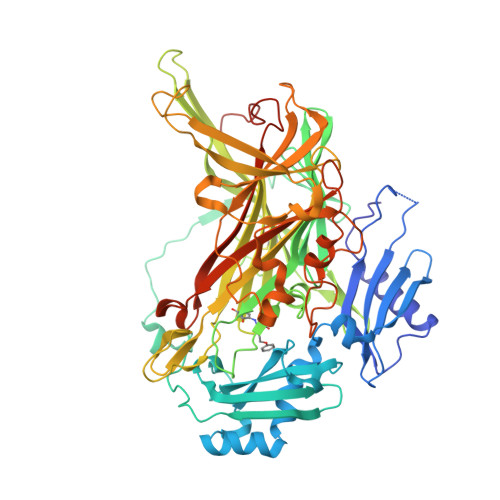 | |
UniProt | |||||
Entity Groups | |||||
| Sequence Clusters | 30% Identity50% Identity70% Identity90% Identity95% Identity100% Identity | ||||
| UniProt Group | P46881 | ||||
Sequence AnnotationsExpand | |||||
Reference Sequence | |||||
| Ligands 4 Unique | |||||
|---|---|---|---|---|---|
| ID | Chains | Name / Formula / InChI Key | 2D Diagram | 3D Interactions | |
| SO4 Download:Ideal Coordinates CCD File | E [auth A], L [auth B], M [auth B], N [auth B] | SULFATE ION O4 S QAOWNCQODCNURD-UHFFFAOYSA-L |  | ||
| GOL Download:Ideal Coordinates CCD File | F [auth A] G [auth A] H [auth A] I [auth A] O [auth B] | GLYCEROL C3 H8 O3 PEDCQBHIVMGVHV-UHFFFAOYSA-N |  | ||
| CU Download:Ideal Coordinates CCD File | C [auth A], J [auth B] | COPPER (II) ION Cu JPVYNHNXODAKFH-UHFFFAOYSA-N |  | ||
| NA Download:Ideal Coordinates CCD File | D [auth A], K [auth B] | SODIUM ION Na FKNQFGJONOIPTF-UHFFFAOYSA-N |  | ||
| Modified Residues 1 Unique | |||||
|---|---|---|---|---|---|
| ID | Chains | Type | Formula | 2D Diagram | Parent |
| P2Q Query on P2Q | A, B | L-PEPTIDE LINKING | C20 H22 N2 O6 |  | TYR |
| Length ( Å ) | Angle ( ˚ ) |
|---|---|
| a = 192.714 | α = 90 |
| b = 62.984 | β = 117.39 |
| c = 158.151 | γ = 90 |
| Software Name | Purpose |
|---|---|
| DENZO | data reduction |
| SCALEPACK | data scaling |
| REFMAC | refinement |
| PDB_EXTRACT | data extraction |
| HKL-2000 | data scaling |


