3KGA | pdb_00003kga

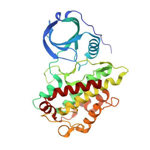
Crystal structure of MAPKAP kinase 2 (MK2) complexed with a potent 3-aminopyrazole ATP site inhibitor


Experimental Data Snapshot

  • Method: X-RAY DIFFRACTION
  • Resolution: 2.55 Å
  • R-Value Free: 
    0.235 (Depositor), 0.234 (DCC) 
  • R-Value Work: 
    0.180 (Depositor), 0.179 (DCC) 
  • R-Value Observed: 
    0.183 (Depositor) 

wwPDB Validation 3D Report Full Report

Validation slider image for 3KGA

Ligand Structure Quality Assessment 


This is version 1.3 of the entry. See complete history. 

Literature

Novel 3-aminopyrazole inhibitors of MK-2 discovered by scaffold hopping strategy.

Velcicky, J., Feifel, R., Hawtin, S., Heng, R., Huppertz, C., Koch, G., Kroemer, M., Moebitz, H., Revesz, L., Scheufler, C., Schlapbach, A.

(2010) Bioorg Med Chem Lett 20: 1293-1297

  • DOI: https://doi.org/10.1016/j.bmcl.2009.10.138
  • Primary Citation Related Structures: 
    3KGA

  • PubMed Abstract: 

    New, selective 3-aminopyrazole based MK2-inhibitors were discovered by scaffold hopping strategy. The new derivatives proved to inhibit intracellular phosphorylation of hsp27 as well as LPS-induced TNFalpha release in cells. In addition, selected derivative 14e also inhibited LPS-induced TNFalpha release in vivo.


  • Organizational Affiliation: 
    • Novartis Institutes for BioMedical Research, CH-4002 Basel, Switzerland. juraj.velcicky@novartis.com

Macromolecule Content 

  • Total Structure Weight: 35.01 kDa 
  • Atom Count: 2,466 
  • Modeled Residue Count: 290 
  • Deposited Residue Count: 299 
  • Unique protein chains: 1

Macromolecules

Find similar proteins by:|  3D Structure
Entity ID: 1
MoleculeChains  Sequence LengthOrganismDetailsImage
MAP kinase-activated protein kinase 2299Homo sapiensMutation(s): 1 
Gene Names: MAPKAPK2
EC: 2.7.11.1
UniProt & NIH Common Fund Data Resources
Find proteins for P49137 (Homo sapiens)
Explore P49137 
Go to UniProtKB:  P49137
PHAROS:  P49137
GTEx:  ENSG00000162889 
Entity Groups
Sequence Clusters30% Identity50% Identity70% Identity90% Identity95% Identity100% Identity
UniProt GroupP49137
Sequence Annotations
Expand
Reference Sequence

Small Molecules

Ligands 2 Unique
IDChains Name / Formula / InChI Key2D Diagram3D Interactions
LX9

Query on LX9


Download:Ideal Coordinates CCD File
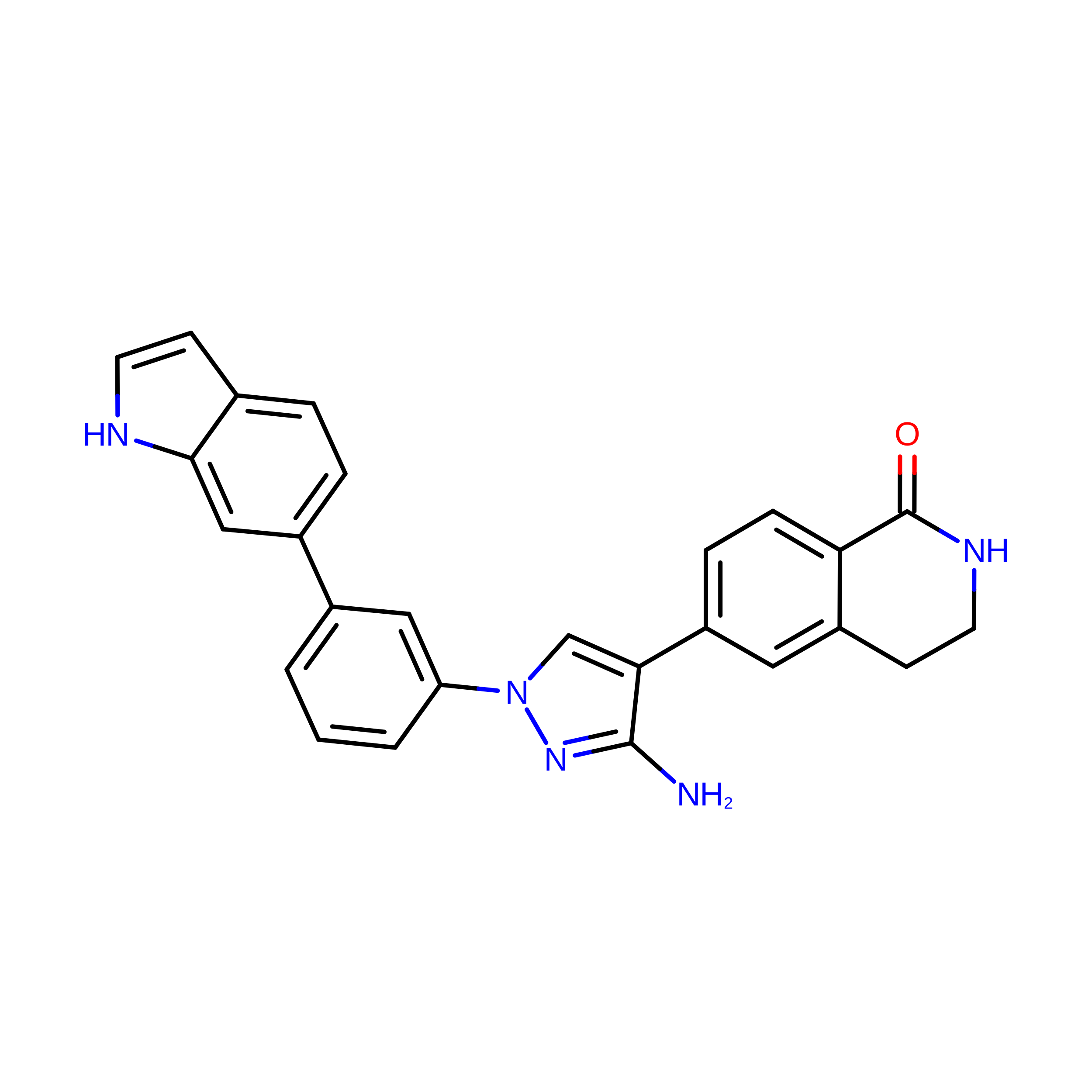
C [auth A]6-{3-amino-1-[3-(1H-indol-6-yl)phenyl]-1H-pyrazol-4-yl}-3,4-dihydroisoquinolin-1(2H)-one
C26 H21 N5 O
ZTKOUSVWVUFWQC-UHFFFAOYSA-N
MG

Query on MG


Download:Ideal Coordinates CCD File
B [auth A]MAGNESIUM ION
Mg
JLVVSXFLKOJNIY-UHFFFAOYSA-N
Binding Affinity Annotations 
IDSourceBinding Affinity
LX9 BindingDB:  3KGA IC50: 61 (nM) from 1 assay(s)

Experimental Data & Validation

Experimental Data

  • Method: X-RAY DIFFRACTION
  • Resolution: 2.55 Å
  • R-Value Free:  0.235 (Depositor), 0.234 (DCC) 
  • R-Value Work:  0.180 (Depositor), 0.179 (DCC) 
  • R-Value Observed: 0.183 (Depositor) 
Space Group: P 63 2 2
Unit Cell:
Length ( Å )Angle ( ˚ )
a = 103.12α = 90
b = 103.12β = 90
c = 165.415γ = 120
Software Package:
Software NamePurpose
REFMACrefinement
XDSdata reduction
XSCALEdata scaling
REFMACphasing

Structure Validation

View Full Validation Report


Ligand Structure Quality Assessment 


Entry History 

Deposition Data

Revision History  (Full details and data files)

  • Version 1.0: 2010-01-26
    Type: Initial release
  • Version 1.1: 2011-07-13
    Changes: Version format compliance
  • Version 1.2: 2021-10-13
    Changes: Database references, Derived calculations
  • Version 1.3: 2024-02-21
    Changes: Data collection