3KAL | pdb_00003kal

Structure of homoglutathione synthetase from Glycine max in closed conformation with homoglutathione, ADP, a sulfate ion, and three magnesium ions bound


Experimental Data Snapshot

  • Method: X-RAY DIFFRACTION
  • Resolution: 1.90 Å
  • R-Value Free: 
    0.250 (Depositor), 0.247 (DCC) 
  • R-Value Work: 
    0.197 (Depositor), 0.198 (DCC) 
  • R-Value Observed: 
    0.200 (Depositor) 

Starting Model: in silico
View more details

wwPDB Validation 3D Report Full Report

Validation slider image for 3KAL

Ligand Structure Quality Assessment 


This is version 1.3 of the entry. See complete history

Literature

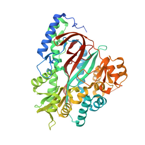
Structural Basis for Evolution of Product Diversity in Soybean Glutathione Biosynthesis.

Galant, A.Arkus, K.A.Zubieta, C.Cahoon, R.E.Jez, J.M.

(2009) Plant Cell 21: 3450-3458

  • DOI: https://doi.org/10.1105/tpc.109.071183
  • Primary Citation Related Structures: 
    3KAJ, 3KAK, 3KAL

  • PubMed Abstract: 

    The redox active peptide glutathione is ubiquitous in nature, but some plants also synthesize glutathione analogs in response to environmental stresses. To understand the evolution of chemical diversity in the closely related enzymes homoglutathione synthetase (hGS) and glutathione synthetase (GS), we determined the structures of soybean (Glycine max) hGS in three states: apoenzyme, bound to gamma-glutamylcysteine (gammaEC), and with hGSH, ADP, and a sulfate ion bound in the active site. Domain movements and rearrangement of active site loops change the structure from an open active site form (apoenzyme and gammaEC complex) to a closed active site form (hGSH*ADP*SO(4)(2-) complex). The structure of hGS shows that two amino acid differences in an active site loop provide extra space to accommodate the longer beta-Ala moiety of hGSH in comparison to the glycinyl group of glutathione. Mutation of either Leu-487 or Pro-488 to an Ala improves catalytic efficiency using Gly, but a double mutation (L487A/P488A) is required to convert the substrate preference of hGS from beta-Ala to Gly. These structures, combined with site-directed mutagenesis, reveal the molecular changes that define the substrate preference of hGS, explain the product diversity within evolutionarily related GS-like enzymes, and reinforce the critical role of active site loops in the adaptation and diversification of enzyme function.


  • Organizational Affiliation
    • Department of Biology, Washington University, St. Louis, Missouri 63130, USA.

Macromolecule Content 

  • Total Structure Weight: 113.93 kDa 
  • Atom Count: 8,571 
  • Modeled Residue Count: 938 
  • Deposited Residue Count: 998 
  • Unique protein chains: 1

Macromolecules

Find similar proteins by:|  3D Structure
Entity ID: 1
MoleculeChains  Sequence LengthOrganismDetailsImage
homoglutathione synthetase
A, B
499Glycine maxMutation(s): 0 
EC: 6.3.2.23 (PDB Primary Data), 6.3.2.3 (UniProt)
UniProt
Find proteins for Q9M426 (Glycine max)
Explore Q9M426 
Go to UniProtKB:  Q9M426
Entity Groups
Sequence Clusters30% Identity50% Identity70% Identity90% Identity95% Identity100% Identity
UniProt GroupQ9M426
Sequence Annotations
Expand
Reference Sequence

Small Molecules

Ligands 4 Unique
IDChains Name / Formula / InChI Key2D Diagram3D Interactions
ADP

Query on ADP


Download:Ideal Coordinates CCD File
C [auth A],
J [auth B]
ADENOSINE-5'-DIPHOSPHATE
C10 H15 N5 O10 P2
XTWYTFMLZFPYCI-KQYNXXCUSA-N
HGS

Query on HGS


Download:Ideal Coordinates CCD File
D [auth A],
I [auth A],
K [auth B],
P [auth B]
D-gamma-glutamyl-L-cysteinyl-beta-alanine
C11 H19 N3 O6 S
HKBNQXMLSMKLJV-RQJHMYQMSA-N
SO4

Query on SO4


Download:Ideal Coordinates CCD File
H [auth A],
O [auth B]
SULFATE ION
O4 S
QAOWNCQODCNURD-UHFFFAOYSA-L
MG

Query on MG


Download:Ideal Coordinates CCD File
E [auth A]
F [auth A]
G [auth A]
L [auth B]
M [auth B]
E [auth A],
F [auth A],
G [auth A],
L [auth B],
M [auth B],
N [auth B]
MAGNESIUM ION
Mg
JLVVSXFLKOJNIY-UHFFFAOYSA-N

Experimental Data & Validation

Experimental Data

  • Method: X-RAY DIFFRACTION
  • Resolution: 1.90 Å
  • R-Value Free:  0.250 (Depositor), 0.247 (DCC) 
  • R-Value Work:  0.197 (Depositor), 0.198 (DCC) 
  • R-Value Observed: 0.200 (Depositor) 
Space Group: P 32
Unit Cell:
Length ( Å )Angle ( ˚ )
a = 115.7α = 90
b = 115.7β = 90
c = 101.76γ = 120
Software Package:
Software NamePurpose
HKL-2000data collection
PHASERphasing
REFMACrefinement
XDSdata reduction
XSCALEdata scaling

Structure Validation

View Full Validation Report


Ligand Structure Quality Assessment 


Entry History 

Deposition Data

Revision History  (Full details and data files)

  • Version 1.0: 2009-12-22
    Type: Initial release
  • Version 1.1: 2011-07-13
    Changes: Version format compliance
  • Version 1.2: 2024-02-21
    Changes: Data collection, Database references, Derived calculations
  • Version 1.3: 2024-04-03
    Changes: Refinement description