3K9X | pdb_00003k9x

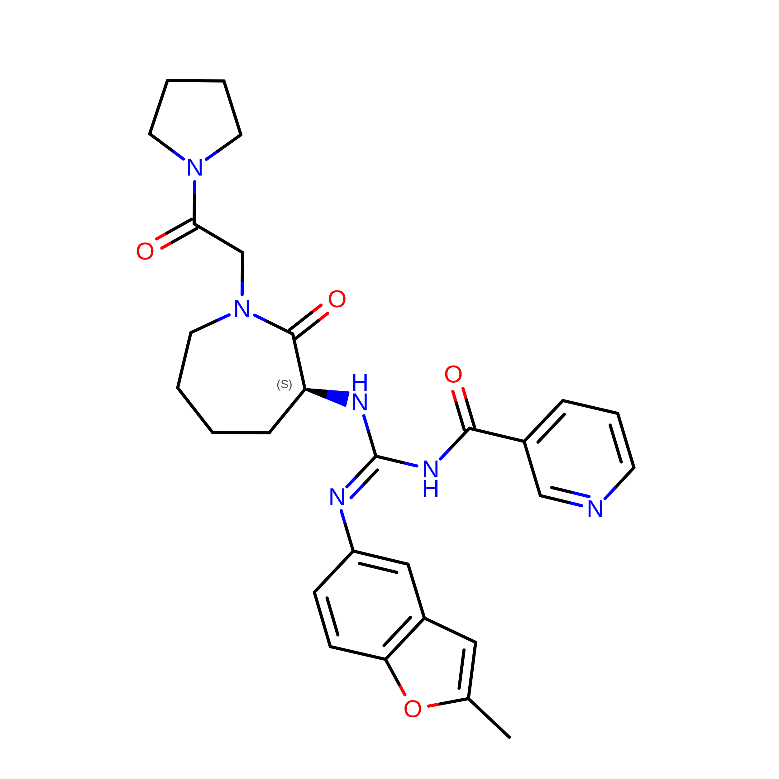
X-ray crystal structure of human fxa in complex with (S)-N-((2-METHYLBENZOFURAN-5-YLAMINO)(2-OXO-1-(2-OXO-2- (PYRROLIDIN-1-YL)ETHYL)AZEPAN-3- YLAMINO)METHYLENE)NICOTINAMIDE


Experimental Data Snapshot

  • Method: X-RAY DIFFRACTION
  • Resolution: 1.90 Å
  • R-Value Free: 
    0.240 (Depositor), 0.238 (DCC) 
  • R-Value Work: 
    0.176 (Depositor), 0.173 (DCC) 
  • R-Value Observed: 
    0.177 (Depositor) 

wwPDB Validation 3D Report Full Report

Validation slider image for 3K9X

Ligand Structure Quality Assessment 


This is version 1.3 of the entry. See complete history. 

Literature

Aroylguanidine-based factor Xa inhibitors: the discovery of BMS-344577

Shi, Y., Li, C., O'Connor, S.P., Zhang, J., Shi, M., Bisaha, S.N., Wang, Y., Sitkoff, D., Pudzianowski, A.T., Huang, C., Klei, H.E., Kish, K., Yanchunas, J., Liu, E.C., Hartl, K.S., Seiler, S.M., Steinbacher, T.E., Schumacher, W.A., Atwal, K.S., Stein, P.D.

(2009) Bioorg Med Chem Lett 19: 6882-6889

  • DOI: https://doi.org/10.1016/j.bmcl.2009.10.084
  • Primary Citation Related Structures: 
    3K9X

  • PubMed Abstract: 

    We report the design and synthesis of a novel class of N,N'-disubstituted aroylguanidine-based lactam derivatives as potent and orally active FXa inhibitors. The structure-activity relationships (SAR) investigation led to the discovery of the nicotinoyl guanidine 22 as a potent FXa inhibitor (FXa IC(50)=4 nM, EC(2xPT)=7 microM). However, the potent CYP3A4 inhibition activity (IC(50)=0.3 microM) of 22 precluded its further development. Detailed analysis of the X-ray crystal structure of compound 22 bound to FXa indicated that the substituent at the 6-position of the nicotinoyl group of 22 would be solvent-exposed, suggesting that efforts to attenuate the unwanted CYP activity could focus at this position without affecting FXa potency significantly. Further SAR studies on the 6-substituted nicotinoyl guanidines resulted in the discovery of 6-(dimethylcarbamoyl) nicotinoyl guanidine 36 (BMS-344577, IC(50)=9 nM, EC(2xPT)=2.5 microM), which was found to be a selective, orally efficacious FXa inhibitor with an excellent in vitro liability profile, favorable pharmacokinetics and pharmacodynamics in animal models.


  • Organizational Affiliation: 
    • Research and Development, Bristol-Myers Squibb Company, PO Box 5400, Princeton, NJ 08543-5400, USA. yan.shi@bms.com

Macromolecule Content 

  • Total Structure Weight: 75.88 kDa 
  • Atom Count: 5,741 
  • Modeled Residue Count: 643 
  • Deposited Residue Count: 664 
  • Unique protein chains: 2

Macromolecules

Find similar proteins by:|  3D Structure
Entity ID: 1
MoleculeChains  Sequence LengthOrganismDetailsImage
PROTEIN (Coagulation factor X)
A, C
94Homo sapiensMutation(s): 0 
EC: 3.4.21.6
UniProt & NIH Common Fund Data Resources
Find proteins for P00742 (Homo sapiens)
Explore P00742 
Go to UniProtKB:  P00742
PHAROS:  P00742
GTEx:  ENSG00000126218 
Entity Groups
Sequence Clusters30% Identity50% Identity70% Identity90% Identity95% Identity100% Identity
UniProt GroupP00742
Sequence Annotations
Expand
Reference Sequence
Find similar proteins by:|  3D Structure
Entity ID: 2
MoleculeChains  Sequence LengthOrganismDetailsImage
PROTEIN (Coagulation factor X)
B, D
238Homo sapiensMutation(s): 0 
EC: 3.4.21.6
UniProt & NIH Common Fund Data Resources
Find proteins for P00742 (Homo sapiens)
Explore P00742 
Go to UniProtKB:  P00742
PHAROS:  P00742
GTEx:  ENSG00000126218 
Entity Groups
Sequence Clusters30% Identity50% Identity70% Identity90% Identity95% Identity100% Identity
UniProt GroupP00742
Sequence Annotations
Expand
Reference Sequence

Small Molecules

Ligands 4 Unique
IDChains Name / Formula / InChI Key2D Diagram3D Interactions
MBM

Query on MBM


Download:Ideal Coordinates CCD File
H [auth B],
M [auth D]
N-{N'-(2-methyl-1-benzofuran-5-yl)-N-[(3S)-2-oxo-1-(2-oxo-2-pyrrolidin-1-ylethyl)azepan-3-yl]carbamimidoyl}pyridine-3-carboxamide
C28 H32 N6 O4
GNYXHJVKEWBHAI-QHCPKHFHSA-N
GOL

Query on GOL


Download:Ideal Coordinates CCD File
E [auth A]
F [auth A]
G [auth A]
K [auth B]
L [auth B]
E [auth A],
F [auth A],
G [auth A],
K [auth B],
L [auth B],
P [auth D]
GLYCEROL
C3 H8 O3
PEDCQBHIVMGVHV-UHFFFAOYSA-N
CA

Query on CA


Download:Ideal Coordinates CCD File
I [auth B],
N [auth D]
CALCIUM ION
Ca
BHPQYMZQTOCNFJ-UHFFFAOYSA-N
NA

Query on NA


Download:Ideal Coordinates CCD File
J [auth B],
O [auth D]
SODIUM ION
Na
FKNQFGJONOIPTF-UHFFFAOYSA-N

Experimental Data & Validation

Experimental Data

  • Method: X-RAY DIFFRACTION
  • Resolution: 1.90 Å
  • R-Value Free:  0.240 (Depositor), 0.238 (DCC) 
  • R-Value Work:  0.176 (Depositor), 0.173 (DCC) 
  • R-Value Observed: 0.177 (Depositor) 
Space Group: P 1 21 1
Unit Cell:
Length ( Å )Angle ( ˚ )
a = 62.263α = 90
b = 77.63β = 102.63
c = 73.732γ = 90
Software Package:
Software NamePurpose
DENZOdata reduction
SCALEPACKdata scaling
AMoREphasing
PHENIXrefinement
PDB_EXTRACTdata extraction

Structure Validation

View Full Validation Report


Ligand Structure Quality Assessment 


Entry History 

Deposition Data

Revision History  (Full details and data files)

  • Version 1.0: 2009-12-15
    Type: Initial release
  • Version 1.1: 2011-07-13
    Changes: Non-polymer description, Version format compliance
  • Version 1.2: 2017-11-01
    Changes: Refinement description
  • Version 1.3: 2024-10-16
    Changes: Data collection, Database references, Derived calculations, Structure summary