3K8A | pdb_00003k8a

Neisseria gonorrhoeae PriB


Experimental Data Snapshot

  • Method: X-RAY DIFFRACTION
  • Resolution: 2.70 Å
  • R-Value Free: 
    0.288 (Depositor), 0.274 (DCC) 
  • R-Value Work: 
    0.243 (Depositor), 0.240 (DCC) 
  • R-Value Observed: 
    0.245 (Depositor) 

Starting Model: experimental
View more details

wwPDB Validation 3D Report Full Report

Validation slider image for 3K8A

This is version 1.3 of the entry. See complete history

Literature

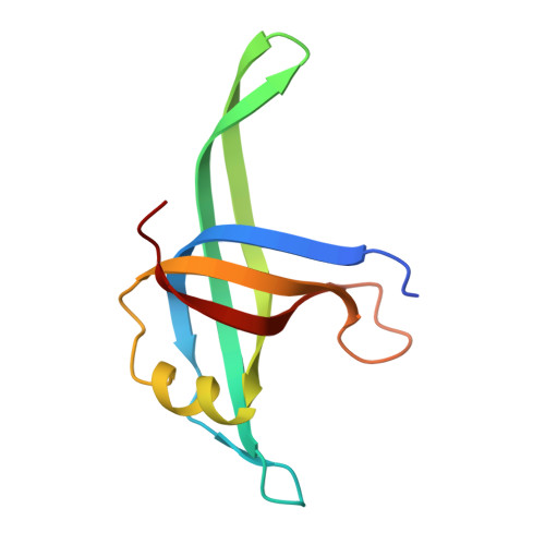
The crystal structure of Neisseria gonorrhoeae PriB reveals mechanistic differences among bacterial DNA replication restart pathways

Dong, J.George, N.P.Duckett, K.L.DeBeer, M.A.Lopper, M.E.

(2010) Nucleic Acids Res 38: 499-509

  • DOI: https://doi.org/10.1093/nar/gkp1031
  • Primary Citation Related Structures: 
    3K8A

  • PubMed Abstract: 

    Reactivation of repaired DNA replication forks is essential for complete duplication of bacterial genomes. However, not all bacteria encode homologs of the well-studied Escherichia coli DNA replication restart primosome proteins, suggesting that there might be distinct mechanistic differences among DNA replication restart pathways in diverse bacteria. Since reactivation of repaired DNA replication forks requires coordinated DNA and protein binding by DNA replication restart primosome proteins, we determined the crystal structure of Neisseria gonorrhoeae PriB at 2.7 A resolution and investigated its ability to physically interact with DNA and PriA helicase. Comparison of the crystal structures of PriB from N. gonorrhoeae and E. coli reveals a well-conserved homodimeric structure consisting of two oligosaccharide/oligonucleotide-binding (OB) folds. In spite of their overall structural similarity, there is significant species variation in the type and distribution of surface amino acid residues. This correlates with striking differences in the affinity with which each PriB homolog binds single-stranded DNA and PriA helicase. These results provide evidence that mechanisms of DNA replication restart are not identical across diverse species and that these pathways have likely become specialized to meet the needs of individual organisms.


  • Organizational Affiliation
    • Department of Chemistry, University of Dayton, 300 College Park, Dayton, OH 45469, USA.

Macromolecule Content 

  • Total Structure Weight: 23.48 kDa 
  • Atom Count: 1,634 
  • Modeled Residue Count: 203 
  • Deposited Residue Count: 206 
  • Unique protein chains: 1

Macromolecules

Find similar proteins by:|  3D Structure
Entity ID: 1
MoleculeChains  Sequence LengthOrganismDetailsImage
Putative primosomal replication protein
A, B
103Neisseria gonorrhoeae FA 1090Mutation(s): 0 
Gene Names: NGO0582prib
UniProt
Find proteins for Q5F924 (Neisseria gonorrhoeae (strain ATCC 700825 / FA 1090))
Explore Q5F924 
Go to UniProtKB:  Q5F924
Entity Groups
Sequence Clusters30% Identity50% Identity70% Identity90% Identity95% Identity100% Identity
UniProt GroupQ5F924
Sequence Annotations
Expand
Reference Sequence

Experimental Data & Validation

Experimental Data

  • Method: X-RAY DIFFRACTION
  • Resolution: 2.70 Å
  • R-Value Free:  0.288 (Depositor), 0.274 (DCC) 
  • R-Value Work:  0.243 (Depositor), 0.240 (DCC) 
  • R-Value Observed: 0.245 (Depositor) 
Space Group: I 41
Unit Cell:
Length ( Å )Angle ( ˚ )
a = 74.24α = 90
b = 74.24β = 90
c = 140.359γ = 90
Software Package:
Software NamePurpose
DENZOdata reduction
SCALEPACKdata scaling
PHASERphasing
REFMACrefinement
PDB_EXTRACTdata extraction
HKL-2000data collection
HKL-2000data reduction
HKL-2000data scaling

Structure Validation

View Full Validation Report


Entry History 

Deposition Data

Revision History  (Full details and data files)

  • Version 1.0: 2010-01-12
    Type: Initial release
  • Version 1.1: 2011-07-13
    Changes: Advisory, Refinement description, Version format compliance
  • Version 1.2: 2017-11-01
    Changes: Refinement description
  • Version 1.3: 2023-09-06
    Changes: Data collection, Database references, Refinement description