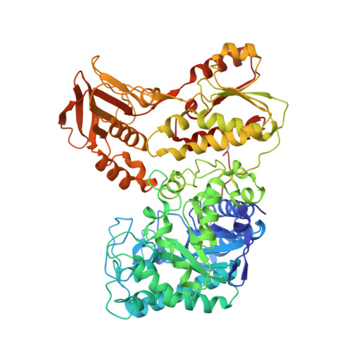
Crystal structure of histamine dehydrogenase from Nocardioides simplex.
Reed, T., Lushington, G.H., Xia, Y., Hirakawa, H., Travis, D.M., Mure, M., Scott, E.E., Limburg, J.(2010) J Biological Chem 285: 25782-25791
- PubMed: 20538584
- DOI: https://doi.org/10.1074/jbc.M109.084301
- Primary Citation of Related Structures:
3K30 - PubMed Abstract:
Histamine dehydrogenase (HADH) isolated from Nocardioides simplex catalyzes the oxidative deamination of histamine to imidazole acetaldehyde. HADH is highly specific for histamine, and we are interested in understanding the recognition mode of histamine in its active site. We describe the first crystal structure of a recombinant form of HADH (HADH) to 2.7-A resolution. HADH is a homodimer, where each 76-kDa subunit contains an iron-sulfur cluster ([4Fe-4S](2+)) and a 6-S-cysteinyl flavin mononucleotide (6-S-Cys-FMN) as redox cofactors. The overall structure of HADH is very similar to that of trimethylamine dehydrogenase (TMADH) from Methylotrophus methylophilus (bacterium W3A1). However, some distinct differences between the structure of HADH and TMADH have been found. Tyr(60), Trp(264), and Trp(355) provide the framework for the "aromatic bowl" that serves as a trimethylamine-binding site in TMADH is comprised of Gln(65), Trp(267), and Asp(358), respectively, in HADH. The surface Tyr(442) that is essential in transferring electrons to electron-transfer flavoprotein (ETF) in TMADH is not conserved in HADH. We use this structure to propose the binding mode for histamine in the active site of HADH through molecular modeling and to compare the interactions to those observed for other histamine-binding proteins whose structures are known.
- Department of Chemistry, The University of Kansas, Lawrence, Kansas 66045, USA.
Organizational Affiliation:


