3JU4 | pdb_00003ju4

Crystal Structure Analysis of EndosialidaseNF at 0.98 A Resolution


Experimental Data Snapshot

  • Method: X-RAY DIFFRACTION
  • Resolution: 0.98 Å
  • R-Value Free: 
    0.133 (Depositor), 0.134 (DCC) 
  • R-Value Work: 
    0.116 (Depositor), 0.122 (DCC) 
  • R-Value Observed: 
    0.116 (Depositor) 

Starting Model: experimental
View more details

wwPDB Validation 3D Report Full Report

Validation slider image for 3JU4

Ligand Structure Quality Assessment 


This is version 1.4 of the entry. See complete history

Literature

Structure analysis of endosialidase NF at 0.98 A resolution.

Schulz, E.C.Neumann, P.Gerardy-Schahn, R.Sheldrick, G.M.Ficner, R.

(2010) Acta Crystallogr D Biol Crystallogr 66: 176-180

  • DOI: https://doi.org/10.1107/S0907444909048720
  • Primary Citation Related Structures: 
    3JU4

  • PubMed Abstract: 

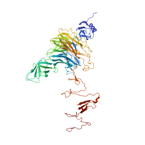
    Endosialidase NF (endoNF) is a bacteriophage-derived endosialidase that specifically degrades alpha-2,8-linked polysialic acid. The structure of a new crystal form of endoNF in complex with sialic acid has been refined at 0.98 A resolution. The 210 kDa homotrimeric multi-domain enzyme displays outstanding stability and resistance to SDS. Even at atomic resolution, only a minor fraction of side chains possess alternative conformations. However, multiple conformations of an active-site residue imply that it has an important catalytic function in the cleavage mechanism of polysialic acid.


  • Organizational Affiliation
    • Abteilung für Molekulare Strukturbiologie, Institut für Mikrobiologie und Genetik, Georg-August-Universität Göttingen, Justus-von-Liebig-Weg 11, 37077 Göttingen, Germany.

Macromolecule Content 

  • Total Structure Weight: 75.05 kDa 
  • Atom Count: 7,145 
  • Modeled Residue Count: 670 
  • Deposited Residue Count: 670 
  • Unique protein chains: 1

Macromolecules

Find similar proteins by:|  3D Structure
Entity ID: 1
MoleculeChains  Sequence LengthOrganismDetailsImage
Endo-N-acetylneuraminidase670Escherichia phage K1FMutation(s): 0 
EC: 3.2.1.129
UniProt
Find proteins for Q04830 (Escherichia phage K1F)
Explore Q04830 
Go to UniProtKB:  Q04830
Entity Groups
Sequence Clusters30% Identity50% Identity70% Identity90% Identity95% Identity100% Identity
UniProt GroupQ04830
Sequence Annotations
Expand
Reference Sequence

Small Molecules

Ligands 3 Unique
IDChains Name / Formula / InChI Key2D Diagram3D Interactions
SLB

Query on SLB


Download:Ideal Coordinates CCD File
B [auth A]N-acetyl-beta-neuraminic acid
C11 H19 N O9
SQVRNKJHWKZAKO-PFQGKNLYSA-N
CL

Query on CL


Download:Ideal Coordinates CCD File
C [auth A],
D [auth A],
E [auth A],
F [auth A],
G [auth A]
CHLORIDE ION
Cl
VEXZGXHMUGYJMC-UHFFFAOYSA-M
NA

Query on NA


Download:Ideal Coordinates CCD File
H [auth A]SODIUM ION
Na
FKNQFGJONOIPTF-UHFFFAOYSA-N

Experimental Data & Validation

Experimental Data

  • Method: X-RAY DIFFRACTION
  • Resolution: 0.98 Å
  • R-Value Free:  0.133 (Depositor), 0.134 (DCC) 
  • R-Value Work:  0.116 (Depositor), 0.122 (DCC) 
  • R-Value Observed: 0.116 (Depositor) 
Space Group: H 3
Unit Cell:
Length ( Å )Angle ( ˚ )
a = 119.04α = 90
b = 119.04β = 90
c = 175.69γ = 120
Software Package:
Software NamePurpose
DNAdata collection
MOLREPphasing
SHELXL-97refinement
XDSdata reduction
XSCALEdata scaling

Structure Validation

View Full Validation Report


Ligand Structure Quality Assessment 


Entry History 

Deposition Data

Revision History  (Full details and data files)

  • Version 1.0: 2010-02-02
    Type: Initial release
  • Version 1.1: 2011-07-13
    Changes: Version format compliance
  • Version 1.2: 2012-04-18
    Changes: Database references
  • Version 1.3: 2020-07-29
    Type: Remediation
    Reason: Carbohydrate remediation
    Changes: Data collection, Database references, Derived calculations, Structure summary
  • Version 1.4: 2023-11-01
    Changes: Data collection, Database references, Refinement description, Structure summary