Single-particle cryoEM analysis at near-atomic resolution from several thousand asymmetric subunits.
Passos, D.O., Lyumkis, D.(2015) J Struct Biol 192: 235-244
- PubMed: 26470814 Search on PubMed
- DOI: https://doi.org/10.1016/j.jsb.2015.10.002
- Primary Citation Related Structures:
3JBS - PubMed Abstract:
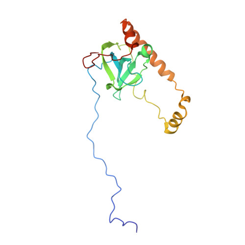
A single-particle cryoEM reconstruction of the large ribosomal subunit from Saccharomyces cerevisiae was obtained from a dataset of ∼75,000 particles. The gold-standard and frequency-limited approaches to single-particle refinement were each independently used to determine orientation parameters for the final reconstruction. Both approaches showed similar resolution curves and nominal resolution values for the 60S dataset, estimated at 2.9 Å. The amount of over-fitting present during frequency-limited refinement was quantitatively analyzed using the high-resolution phase-randomization test, and the results showed no apparent over-fitting. The number of asymmetric subunits required to reach specific resolutions was subsequently analyzed by refining subsets of the data in an ab initio manner. With our data collection and processing strategies, sub-nanometer resolution was obtained with ∼200 asymmetric subunits (or, equivalently for the ribosomal subunit, particles). Resolutions of 5.6 Å, 4.5 Å, and 3.8 Å were reached with ∼1000, ∼1600, and ∼5000 asymmetric subunits, respectively. At these resolutions, one would expect to detect alpha-helical pitch, separation of beta-strands, and separation of Cα atoms, respectively. Using this map, together with strategies for ab initio model building and model refinement, we built a region of the ribosomal protein eL6, which was missing in previous models of the yeast ribosome. The relevance for more routine high-resolution structure determination is discussed.
- Laboratory of Genetics and Helmsley Center for Genomic Medicine, The Salk Institute for Biological Studies, 10010 North Torrey Pines Road, La Jolla, CA 92037, United States.
Organizational Affiliation: