3J8D

Cryoelectron microscopy of dengue-Fab E104 complex at pH 5.5


Experimental Data Snapshot

  • Method: ELECTRON MICROSCOPY
  • Resolution: 26.0 Å
  • Aggregation State: PARTICLE 
  • Reconstruction Method: SINGLE PARTICLE 

Starting Models: experimental
View more details

wwPDB Validation   3D Report Full Report


This is version 1.4 of the entry. See complete history


Literature

Structure of Acidic pH Dengue Virus Showing the Fusogenic Glycoprotein Trimers.

Zhang, X.Sheng, J.Austin, S.K.Hoornweg, T.E.Smit, J.M.Kuhn, R.J.Diamond, M.S.Rossmann, M.G.

(2015) J Virol 89: 743-750

  • DOI: https://doi.org/10.1128/JVI.02411-14
  • Primary Citation of Related Structures:  
    3J8D

  • PubMed Abstract: 

    Flaviviruses undergo large conformational changes during their life cycle. Under acidic pH conditions, the mature virus forms transient fusogenic trimers of E glycoproteins that engage the lipid membrane in host cells to initiate viral fusion and nucleocapsid penetration into the cytoplasm. However, the dynamic nature of the fusogenic trimer has made the determination of its structure a challenge. Here we have used Fab fragments of the neutralizing antibody DV2-E104 to stop the conformational change of dengue virus at an intermediate stage of the fusion process. Using cryo-electron microscopy, we show that in this intermediate stage, the E glycoproteins form 60 trimers that are similar to the predicted "open" fusogenic trimer. The structure of a dengue virus has been captured during the formation of fusogenic trimers. This was accomplished by binding Fab fragments of the neutralizing antibody DV2-E104 to the virus at neutral pH and then decreasing the pH to 5.5. These trimers had an "open" conformation, which is distinct from the "closed" conformation of postfusion trimers. Only two of the three E proteins within each spike are bound by a Fab molecule at domain III. Steric hindrance around the icosahedral 3-fold axes prevents binding of a Fab to the third domain III of each E protein spike. Binding of the DV2-E104 Fab fragments prevents domain III from rotating by about 130° to the postfusion orientation and thus precludes the stem region from "zipping" together the three E proteins along the domain II boundaries into the "closed" postfusion conformation, thus inhibiting fusion.


  • Organizational Affiliation

    Department of Biological Sciences, Purdue University, West Lafayette, Indiana, USA.


Macromolecules
Find similar proteins by:  (by identity cutoff)  |  3D Structure
Entity ID: 1
MoleculeChains Sequence LengthOrganismDetailsImage
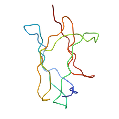
glycoprotein DIIIA [auth B],
D [auth F]
97Dengue virus 2 Thailand/16681/84Mutation(s): 0 
UniProt
Find proteins for P17763 (Dengue virus type 1 (strain Nauru/West Pac/1974))
Explore P17763 
Go to UniProtKB:  P17763
Entity Groups  
Sequence Clusters30% Identity50% Identity70% Identity90% Identity95% Identity100% Identity
UniProt GroupP17763
Sequence Annotations
Expand
  • Reference Sequence
Find similar proteins by:  (by identity cutoff)  |  3D Structure
Entity ID: 2
MoleculeChains Sequence LengthOrganismDetailsImage
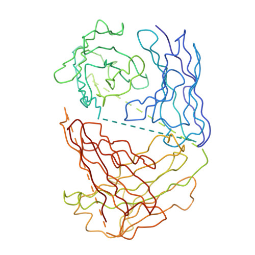
antibody E111 Fab fragmentB [auth A],
C [auth D]
425Mus musculusMutation(s): 0 
Entity Groups  
Sequence Clusters30% Identity50% Identity70% Identity90% Identity95% Identity100% Identity
Sequence Annotations
Expand
  • Reference Sequence
Find similar proteins by:  (by identity cutoff)  |  3D Structure
Entity ID: 3
MoleculeChains Sequence LengthOrganismDetailsImage
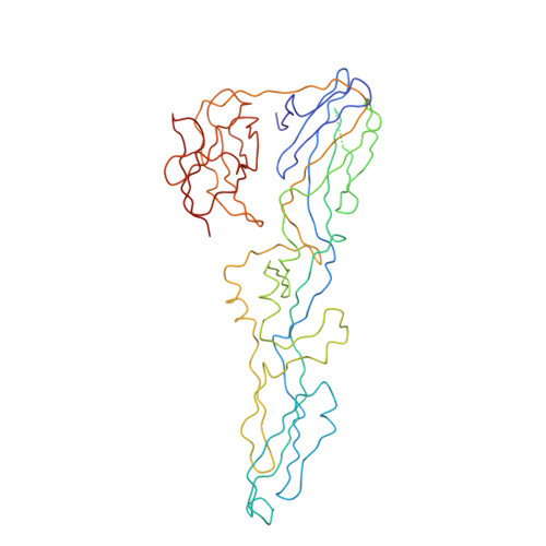
Envelope protein EE [auth G],
F [auth H],
G [auth I]
394Dengue virus 2 Thailand/16681/84Mutation(s): 0 
UniProt
Find proteins for P29990 (Dengue virus type 2 (strain Thailand/16681/1984))
Explore P29990 
Go to UniProtKB:  P29990
Entity Groups  
Sequence Clusters30% Identity50% Identity70% Identity90% Identity95% Identity100% Identity
UniProt GroupP29990
Sequence Annotations
Expand
  • Reference Sequence
Experimental Data & Validation

Experimental Data

  • Method: ELECTRON MICROSCOPY
  • Resolution: 26.0 Å
  • Aggregation State: PARTICLE 
  • Reconstruction Method: SINGLE PARTICLE 
EM Software:
TaskSoftware PackageVersion
RECONSTRUCTIONEMAN

Structure Validation

View Full Validation Report


Entry History 

Deposition Data

Revision History  (Full details and data files)

  • Version 1.0: 2014-11-12
    Type: Initial release
  • Version 1.1: 2014-12-24
    Changes: Database references
  • Version 1.2: 2017-07-26
    Changes: Source and taxonomy
  • Version 1.3: 2018-07-18
    Changes: Data collection
  • Version 1.4: 2024-02-21
    Changes: Data collection, Database references, Derived calculations, Refinement description