3J2Q | pdb_00003j2q

Model of membrane-bound factor VIII organized in 2D crystals


Experimental Data Snapshot

  • Method: ELECTRON CRYSTALLOGRAPHY
  • R-Value Work: 
    0.426 (DCC) 

wwPDB Validation 3D Report Full Report

Validation slider image for 3J2Q

This is version 2.1 of the entry. See complete history

Literature

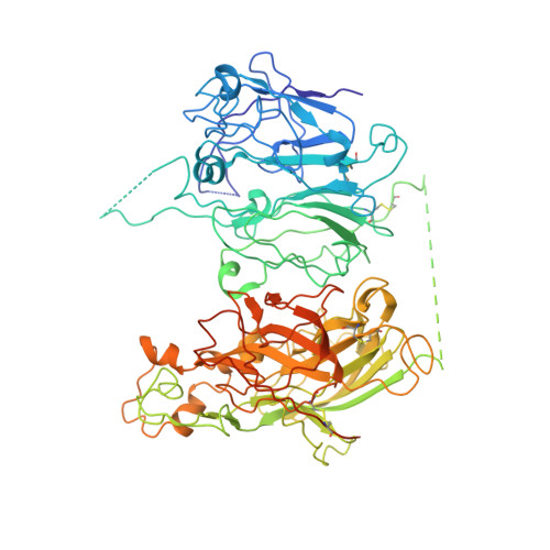
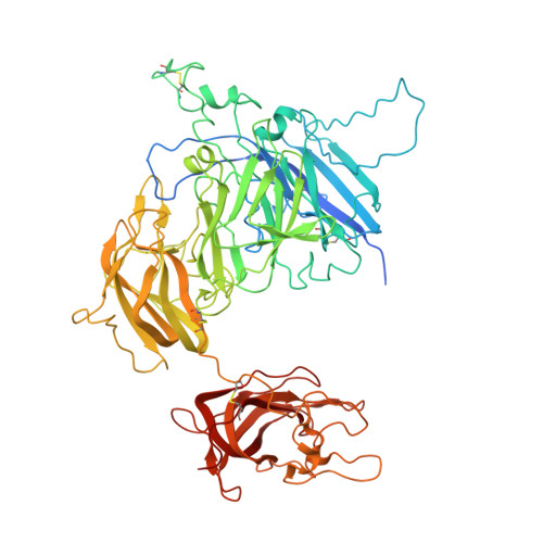
Domain organization of membrane-bound factor VIII.

Stoilova-McPhie, S.Lynch, G.C.Ludtke, S.Pettitt, B.M.

(2013) Biopolymers 99: 448-459

  • DOI: https://doi.org/10.1002/bip.22199
  • Primary Citation Related Structures: 
    3J2Q, 3J2S

  • PubMed Abstract: 

    Factor VIII (FVIII) is the blood coagulation protein which when defective or deficient causes for hemophilia A, a severe hereditary bleeding disorder. Activated FVIII (FVIIIa) is the cofactor to the serine protease factor IXa (FIXa) within the membrane-bound Tenase complex, responsible for amplifying its proteolytic activity more than 100,000 times, necessary for normal clot formation. FVIII is composed of two noncovalently linked peptide chains: a light chain (LC) holding the membrane interaction sites and a heavy chain (HC) holding the main FIXa interaction sites. The interplay between the light and heavy chains (HCs) in the membrane-bound state is critical for the biological efficiency of FVIII. Here, we present our cryo-electron microscopy (EM) and structure analysis studies of human FVIII-LC, when helically assembled onto negatively charged single lipid bilayer nanotubes. The resolved FVIII-LC membrane-bound structure supports aspects of our previously proposed FVIII structure from membrane-bound two-dimensional (2D) crystals, such as only the C2 domain interacts directly with the membrane. The LC is oriented differently in the FVIII membrane-bound helical and 2D crystal structures based on EM data, and the existing X-ray structures. This flexibility of the FVIII-LC domain organization in different states is discussed in the light of the FVIIIa-FIXa complex assembly and function.


  • Organizational Affiliation
    • Department of Neuroscience and Cell Biology, University of Texas Medical Branch, Galveston, TX, USA. svmcphie@utmb.edu

Macromolecule Content 

  • Total Structure Weight: 167.25 kDa 
  • Atom Count: 10,317 
  • Modeled Residue Count: 1,261 
  • Deposited Residue Count: 1,438 
  • Unique protein chains: 2

Macromolecules

Find similar proteins by:|  3D Structure
Entity ID: 1
MoleculeChains  Sequence LengthOrganismDetailsImage
Coagulation factor VIII heavy chain754Homo sapiensMutation(s): 0 
Gene Names: F8F8C
Membrane Entity: Yes 
UniProt & NIH Common Fund Data Resources
Find proteins for P00451 (Homo sapiens)
Explore P00451 
Go to UniProtKB:  P00451
PHAROS:  P00451
GTEx:  ENSG00000185010 
Entity Groups
Sequence Clusters30% Identity50% Identity70% Identity90% Identity95% Identity100% Identity
UniProt GroupP00451
Glycosylation
Glycosylation Sites: 1Go to GlyGen: P00451-1
Sequence Annotations
Expand
Reference Sequence
Find similar proteins by:|  3D Structure
Entity ID: 2
MoleculeChains  Sequence LengthOrganismDetailsImage
Coagulation factor VIII light chain684Homo sapiensMutation(s): 0 
Gene Names: F8F8C
Membrane Entity: Yes 
UniProt & NIH Common Fund Data Resources
Find proteins for P00451 (Homo sapiens)
Explore P00451 
Go to UniProtKB:  P00451
PHAROS:  P00451
GTEx:  ENSG00000185010 
Entity Groups
Sequence Clusters30% Identity50% Identity70% Identity90% Identity95% Identity100% Identity
UniProt GroupP00451
Glycosylation
Glycosylation Sites: 2Go to GlyGen: P00451-1
Sequence Annotations
Expand
Reference Sequence

Oligosaccharides

Help  
Entity ID: 3
MoleculeChains Length2D Diagram GlycosylationD Interactions
2-acetamido-2-deoxy-beta-D-glucopyranose-(1-4)-2-acetamido-2-deoxy-beta-D-glucopyranose
C
2N-Glycosylation
Glycosylation Resources
GlyTouCan: G42666HT
GlyCosmos: G42666HT
GlyGen: G42666HT
Entity ID: 4
MoleculeChains Length2D Diagram GlycosylationD Interactions
alpha-D-mannopyranose-(1-3)-[beta-D-mannopyranose-(1-6)]beta-D-mannopyranose-(1-4)-2-acetamido-2-deoxy-beta-D-glucopyranose
D
4N/A
Glycosylation Resources
GlyTouCan: G11348GW
GlyCosmos: G11348GW
GlyGen: G11348GW

Experimental Data & Validation

Experimental Data

  • Method: ELECTRON CRYSTALLOGRAPHY
  • R-Value Work:  0.426 (DCC) 
Space Group: P 1
Unit Cell:
Length ( Å )Angle ( ˚ )
a = 81α = 67
b = 71β = 60
c = 100γ = 65

Structure Validation

View Full Validation Report


Entry History 

Deposition Data

Revision History  (Full details and data files)

  • Version 1.0: 2013-09-11
    Type: Initial release
  • Version 2.0: 2020-07-29
    Type: Remediation
    Reason: Carbohydrate remediation
    Changes: Advisory, Atomic model, Author supporting evidence, Data collection, Database references, Derived calculations, Structure summary
  • Version 2.1: 2024-11-20
    Changes: Data collection, Database references, Structure summary