3J1Q | pdb_00003j1q

Structure of AAV-DJ, a Retargeted Gene Therapy Vector: Cryo-Electron Microscopy at 4.5A resolution


Experimental Data Snapshot

  • Method: ELECTRON MICROSCOPY
  • Resolution: 4.50 Å
  • Aggregation State: PARTICLE 
  • Reconstruction Method: SINGLE PARTICLE 

Starting Model: experimental
View more details

wwPDB Validation 3D Report Full Report

Validation slider image for 3J1Q

This is version 1.3 of the entry. See complete history

Literature

Structure of AAV-DJ, a retargeted gene therapy vector: cryo-electron microscopy at 4.5 A resolution.

Lerch, T.F.O'Donnell, J.K.Meyer, N.L.Xie, Q.Taylor, K.A.Stagg, S.M.Chapman, M.S.

(2012) Structure 20: 1310-1320

  • DOI: https://doi.org/10.1016/j.str.2012.05.004
  • Primary Citation Related Structures: 
    3J1Q

  • PubMed Abstract: 

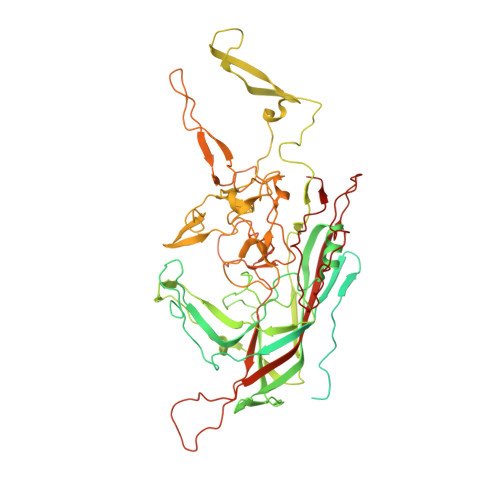
    AAV-DJ, a leading candidate vector for liver gene therapy, was created through random homologous recombination followed by directed evolution, selecting for in vivo liver tropism and resistance to in vitro immune neutralization. Here, the 4.5 Å resolution cryo-EM structure is determined for the engineered AAV vector, revealing structural features that illuminate its phenotype. The heparan sulfate receptor-binding site is little changed from AAV-2, and heparin-binding affinity is similar. A loop that is antigenic in other serotypes has a unique conformation in AAV-DJ that would conflict with the binding of an AAV-2 neutralizing monoclonal antibody. This is consistent with increased resistance to neutralization by human polyclonal sera, raising the possibility that changed tropism may be a secondary effect of altered immune interactions. The reconstruction exemplifies analysis of fine structural changes and the potential of cryo-EM, in favorable cases, to characterize mutant or ligand-bound complexes.


  • Organizational Affiliation
    • Department of Biochemistry and Molecular Biology, School of Medicine, Oregon Health and Science University, Portland, OR 97239, USA.

Macromolecule Content 

  • Total Structure Weight: 82.01 kDa 
  • Atom Count: 4,136 
  • Modeled Residue Count: 517 
  • Deposited Residue Count: 737 
  • Unique protein chains: 1

Macromolecules

Find similar proteins by:|  3D Structure
Entity ID: 1
MoleculeChains  Sequence LengthOrganismDetailsImage
Adeno-associated virus DJ737Adeno-associated virusMutation(s): 0 
Gene Names: Capsid
UniProt
Find proteins for Q6JC41 (Adeno-associated virus)
Explore Q6JC41 
Go to UniProtKB:  Q6JC41
Entity Groups
Sequence Clusters30% Identity50% Identity70% Identity90% Identity95% Identity100% Identity
UniProt GroupQ6JC41
Sequence Annotations
Expand
Reference Sequence

Experimental Data & Validation

Experimental Data

  • Method: ELECTRON MICROSCOPY
  • Resolution: 4.50 Å
  • Aggregation State: PARTICLE 
  • Reconstruction Method: SINGLE PARTICLE 
EM Software:
TaskSoftware PackageVersion
RECONSTRUCTIONFREALIGN

Structure Validation

View Full Validation Report


Entry History 

Deposition Data

Revision History  (Full details and data files)

  • Version 1.0: 2012-08-22
    Type: Initial release
  • Version 1.1: 2012-10-24
    Changes: Database references
  • Version 1.2: 2018-07-18
    Changes: Data collection
  • Version 1.3: 2024-02-21
    Changes: Data collection, Database references, Derived calculations, Refinement description