Structure of a type III secretion needle at 7-A resolution provides insights into its assembly and signaling mechanisms.
Fujii, T., Cheung, M., Blanco, A., Kato, T., Blocker, A.J., Namba, K.(2012) Proc Natl Acad Sci U S A 109: 4461-4466
- PubMed: 22388746 Search on PubMedSearch on PubMed Central
- DOI: https://doi.org/10.1073/pnas.1116126109
- Primary Citation Related Structures:
3J0R - PubMed Abstract:
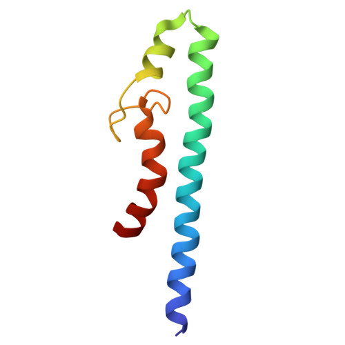
Type III secretion systems of Gram-negative bacteria form injection devices that deliver effector proteins into eukaryotic cells during infection. They span both bacterial membranes and the extracellular space to connect with the host cell plasma membrane. Their extracellular portion is a needle-like, hollow tube that serves as a secretion conduit for effector proteins. The needle of Shigella flexneri is approximately 50-nm long and 7-nm thick and is made by the helical assembly of one protein, MxiH. We provide a 7-Å resolution 3D image reconstruction of the Shigella needle by electron cryomicroscopy, which resolves α-helices and a β-hairpin that has never been observed in the crystal and solution structures of needle proteins, including MxiH. An atomic model of the needle based on the 3D-density map, in comparison with that of the bacterial-flagellar filament, provides insights into how such a thin tubular structure is stably assembled by intricate intermolecular interactions. The map also illuminates how the needle-length control protein functions as a ruler within the central channel during export of MxiH for assembly at the distal end of the needle, and how the secretion-activation signal may be transduced through a conformational change of the needle upon host-cell contact.
- Graduate School of Frontier Biosciences, Osaka University, 1-3 Yamadaoka, Suita, Osaka 565-0871, Japan.
Organizational Affiliation: