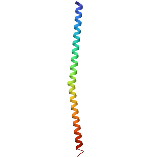
3IYV

Clathrin D6 coat as full-length Triskelions


Experimental Data Snapshot

  • Method: ELECTRON MICROSCOPY
  • Resolution: 7.90 Å
  • Aggregation State: PARTICLE 
  • Reconstruction Method: SINGLE PARTICLE 

Starting Models: experimental
View more details

wwPDB Validation   3D Report Full Report


This is version 1.4 of the entry. See complete history


Literature

Molecular model for a complete clathrin lattice from electron cryomicroscopy.

Fotin, A.Cheng, Y.Sliz, P.Grigorieff, N.Harrison, S.C.Kirchhausen, T.Walz, T.

(2004) Nature 432: 573-579

  • DOI: https://doi.org/10.1038/nature03079
  • Primary Citation of Related Structures:  
    1XI4, 3IYV

  • PubMed Abstract: 

    Clathrin-coated vesicles are important vehicles of membrane traffic in cells. We report the structure of a clathrin lattice at subnanometre resolution, obtained from electron cryomicroscopy of coats assembled in vitro. We trace most of the 1,675-residue clathrin heavy chain by fitting known crystal structures of two segments, and homology models of the rest, into the electron microscopy density map. We also define the position of the central helical segment of the light chain. A helical tripod, the carboxy-terminal parts of three heavy chains, projects inward from the vertex of each three-legged clathrin triskelion, linking that vertex to 'ankles' of triskelions centred two vertices away. Analysis of coats with distinct diameters shows an invariant pattern of contacts in the neighbourhood of each vertex, with more variable interactions along the extended parts of the triskelion 'legs'. These invariant local interactions appear to stabilize the lattice, allowing assembly and uncoating to be controlled by events at a few specific sites.


  • Organizational Affiliation

    Biophysics Graduate Program, Department of Cell Biology, Harvard Medical School, 240 Longwood Avenue, Boston, Massachusetts 02115, USA.


Macromolecules
Find similar proteins by:  (by identity cutoff)  |  3D Structure
Entity ID: 1
MoleculeChains Sequence LengthOrganismDetailsImage
Clathrin heavy chain1,630Bos taurusMutation(s): 0 
UniProt
Find proteins for P49951 (Bos taurus)
Explore P49951 
Go to UniProtKB:  P49951
Entity Groups  
Sequence Clusters30% Identity50% Identity70% Identity90% Identity95% Identity100% Identity
UniProt GroupP49951
Sequence Annotations
Expand
  • Reference Sequence
Find similar proteins by:  (by identity cutoff)  |  3D Structure
Entity ID: 2
MoleculeChains Sequence LengthOrganismDetailsImage
Clathrin light chain A70Bos taurusMutation(s): 0 
UniProt
Find proteins for P04973 (Bos taurus)
Explore P04973 
Go to UniProtKB:  P04973
Entity Groups  
Sequence Clusters30% Identity50% Identity70% Identity90% Identity95% Identity100% Identity
UniProt GroupP04973
Sequence Annotations
Expand
  • Reference Sequence
Experimental Data & Validation

Experimental Data

  • Method: ELECTRON MICROSCOPY
  • Resolution: 7.90 Å
  • Aggregation State: PARTICLE 
  • Reconstruction Method: SINGLE PARTICLE 
EM Software:
TaskSoftware PackageVersion
RECONSTRUCTIONFREALIGN
RECONSTRUCTIONIMAGIC

Structure Validation

View Full Validation Report


Entry History 

Deposition Data

Revision History  (Full details and data files)

  • Version 1.0: 2010-07-21
    Type: Initial release
  • Version 1.1: 2011-07-13
    Changes: Version format compliance
  • Version 1.2: 2018-07-18
    Changes: Data collection
  • Version 1.3: 2019-11-06
    Changes: Data collection, Other
  • Version 1.4: 2024-02-21
    Changes: Data collection, Database references, Derived calculations, Refinement description