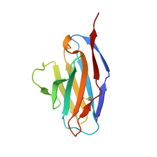
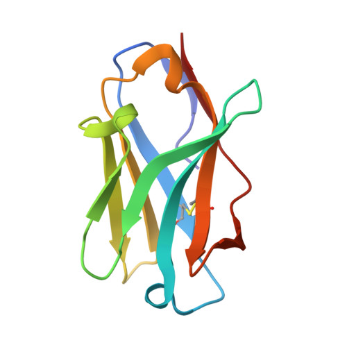
Structural comparison of different antibodies interacting with parvovirus capsids
Hafenstein, S., Bowman, V.D., Sun, T., Nelson, C.D., Palermo, L.M., Chipman, P.R., Battisti, A.J., Parrish, C.R., Rossmann, M.G.(2009) J Virol 83: 5556-5566
- PubMed: 19321620 Search on PubMedSearch on PubMed Central
- DOI: https://doi.org/10.1128/JVI.02532-08
- Primary Citation Related Structures:
3GK8, 3IY0, 3IY1, 3IY2, 3IY3, 3IY4, 3IY5, 3IY6, 3IY7 - PubMed Abstract:
The structures of canine parvovirus (CPV) and feline parvovirus (FPV) complexed with antibody fragments from eight different neutralizing monoclonal antibodies were determined by cryo-electron microscopy (cryoEM) reconstruction to resolutions varying from 8.5 to 18 A. The crystal structure of one of the Fab molecules and the sequence of the variable domain for each of the Fab molecules have been determined. The structures of Fab fragments not determined crystallographically were predicted by homology modeling according to the amino acid sequence. Fitting of the Fab and virus structures into the cryoEM densities identified the footprints of each antibody on the viral surface. As anticipated from earlier analyses, the Fab binding sites are directed to two epitopes, A and B. The A site is on an exposed part of the surface near an icosahedral threefold axis, whereas the B site is about equidistant from the surrounding five-, three-, and twofold axes. One antibody directed to the A site binds CPV but not FPV. Two of the antibodies directed to the B site neutralize the virus as Fab fragments. The differences in antibody properties have been linked to the amino acids within the antibody footprints, the position of the binding site relative to the icosahedral symmetry elements, and the orientation of the Fab structure relative to the surface of the virus. Most of the exposed surface area was antigenic, although each of the antibodies had a common area of overlap that coincided with the positions of the previously mapped escape mutations.
- Department of Biological Sciences, Purdue University, West Lafayette, IN 47907-2054, USA.
Organizational Affiliation: