3IXO | pdb_00003ixo

Crystal Structure of uncomplexed HIV_1 Protease Subtype A


Experimental Data Snapshot

  • Method: X-RAY DIFFRACTION
  • Resolution: 1.70 Å
  • R-Value Free: 
    0.244 (Depositor), 0.245 (DCC) 
  • R-Value Work: 
    0.205 (Depositor), 0.207 (DCC) 
  • R-Value Observed: 
    0.205 (Depositor) 

Starting Model: experimental
View more details

wwPDB Validation 3D Report Full Report

Validation slider image for 3IXO

This is version 1.3 of the entry. See complete history

Literature

Structure of the unbound form of HIV-1 subtype A protease: comparison with unbound forms of proteases from other HIV subtypes.

Robbins, A.H.Coman, R.M.Bracho-Sanchez, E.Fernandez, M.A.Gilliland, C.T.Li, M.Agbandje-McKenna, M.Wlodawer, A.Dunn, B.M.McKenna, R.

(2010) Acta Crystallogr D Biol Crystallogr 66: 233-242

  • DOI: https://doi.org/10.1107/S0907444909054298
  • Primary Citation Related Structures: 
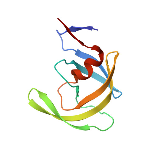
    3IXO

  • PubMed Abstract: 

    The crystal structure of the unbound form of HIV-1 subtype A protease (PR) has been determined to 1.7 A resolution and refined as a homodimer in the hexagonal space group P6(1) to an R(cryst) of 20.5%. The structure is similar in overall shape and fold to the previously determined subtype B, C and F PRs. The major differences lie in the conformation of the flap region. The flaps in the crystal structures of the unbound subtype B and C PRs, which were crystallized in tetragonal space groups, are either semi-open or wide open. In the present structure of subtype A PR the flaps are found in the closed position, a conformation that would be more anticipated in the structure of HIV protease complexed with an inhibitor. The amino-acid differences between the subtypes and their respective crystal space groups are discussed in terms of the differences in the flap conformations.


  • Organizational Affiliation
    • Department of Biochemistry and Molecular Biology, University of Florida, Gainesville, FL 32610, USA.

Macromolecule Content 

  • Total Structure Weight: 21.54 kDa 
  • Atom Count: 1,617 
  • Modeled Residue Count: 198 
  • Deposited Residue Count: 198 
  • Unique protein chains: 1

Macromolecules

Find similar proteins by:|  3D Structure
Entity ID: 1
MoleculeChains  Sequence LengthOrganismDetailsImage
HIV-1 protease
A, B
99HIV-1 M:AMutation(s): 2 
Gene Names: pol
UniProt
Find proteins for P04588 (Human immunodeficiency virus type 1 group M subtype A (isolate MAL))
Explore P04588 
Go to UniProtKB:  P04588
Entity Groups
Sequence Clusters30% Identity50% Identity70% Identity90% Identity95% Identity100% Identity
UniProt GroupP04588
Sequence Annotations
Expand
Reference Sequence

Experimental Data & Validation

Experimental Data

  • Method: X-RAY DIFFRACTION
  • Resolution: 1.70 Å
  • R-Value Free:  0.244 (Depositor), 0.245 (DCC) 
  • R-Value Work:  0.205 (Depositor), 0.207 (DCC) 
  • R-Value Observed: 0.205 (Depositor) 
Space Group: P 61
Unit Cell:
Length ( Å )Angle ( ˚ )
a = 62.207α = 90
b = 62.207β = 90
c = 82.47γ = 120
Software Package:
Software NamePurpose
CNSrefinement
HKL-2000data reduction
HKL-2000data scaling
CNSphasing

Structure Validation

View Full Validation Report


Entry History 

Deposition Data

Revision History  (Full details and data files)

  • Version 1.0: 2010-03-02
    Type: Initial release
  • Version 1.1: 2011-07-13
    Changes: Version format compliance
  • Version 1.2: 2012-03-28
    Changes: Database references
  • Version 1.3: 2023-09-06
    Changes: Data collection, Database references, Refinement description