Design and Synthesis of Potent and Selective BACE-1 Inhibitors.
Bjorklund, C., Oscarson, S., Benkestock, K., Borkakoti, N., Jansson, K., Lindberg, J., Vrang, L., Hallberg, A., Rosenquist, A., Samuelsson, B.(2010) J Med Chem 53: 1458-1464
- PubMed: 20128595 Search on PubMed
- DOI: https://doi.org/10.1021/jm901168f
- Primary Citation Related Structures:
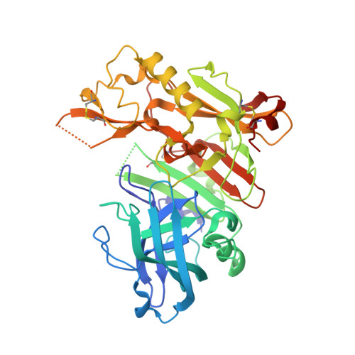
3IXJ - PubMed Abstract:
Highly potent BACE-1 protease inhibitors have been developed from an inhibitors containing a hydroxyethylene (HE) core displaying aryloxymethyl or benzyloxymethyl P1 side chain and a methoxy P1' side chain. The target molecules were synthesized in good overall yields from chiral carbohydrate starting materials. The inhibitors show high BACE-1 potency and good selectivity against cathepsin D, where the most potent inhibitor furnishes BACE-1 K(i) << 1 nM and displays >1000-fold selectivity over cathepsin D.
- Department of Organic Chemistry, Arrhenius Laboratory, Stockholm University, SE-106 91 Stockholm, Sweden.
Organizational Affiliation: