3ITN | pdb_00003itn

Crystal structure of Pseudo-activated Procaspase-3


Experimental Data Snapshot

  • Method: X-RAY DIFFRACTION
  • Resolution: 1.63 Å
  • R-Value Free: 
    0.224 (Depositor), 0.220 (DCC) 
  • R-Value Work: 
    0.199 (Depositor), 0.207 (DCC) 
  • R-Value Observed: 
    0.199 (Depositor) 

Starting Model: experimental
View more details

wwPDB Validation 3D Report Full Report

Validation slider image for 3ITN

This is version 1.4 of the entry. See complete history

Literature

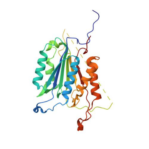
A constitutively active and uninhibitable caspase-3 zymogen efficiently induces apoptosis.

Walters, J.Pop, C.Scott, F.L.Drag, M.Swartz, P.Mattos, C.Salvesen, G.S.Clark, A.C.

(2009) Biochem J 424: 335-345

  • DOI: https://doi.org/10.1042/BJ20090825
  • Primary Citation Related Structures: 
    3ITN

  • PubMed Abstract: 

    The caspase-3 zymogen has essentially zero activity until it is cleaved by initiator caspases during apoptosis. However, a mutation of V266E in the dimer interface activates the protease in the absence of chain cleavage. We show that low concentrations of the pseudo-activated procaspase-3 kill mammalian cells rapidly and, importantly, this protein is not cleaved nor is it inhibited efficiently by the endogenous regulator XIAP (X-linked inhibitor of apoptosis). The 1.63 A (1 A = 0.1 nm) structure of the variant demonstrates that the mutation is accommodated at the dimer interface to generate an enzyme with substantially the same activity and specificity as wild-type caspase-3. Structural modelling predicts that the interface mutation prevents the intersubunit linker from binding in the dimer interface, allowing the active sites to form in the procaspase in the absence of cleavage. The direct activation of procaspase-3 through a conformational switch rather than by chain cleavage may lead to novel therapeutic strategies for inducing cell death.


  • Organizational Affiliation
    • Department of Molecular and Structural Biochemistry, North Carolina State University, Raleigh, NC 27695, USA.

Macromolecule Content 

  • Total Structure Weight: 29.17 kDa 
  • Atom Count: 2,199 
  • Modeled Residue Count: 246 
  • Deposited Residue Count: 256 
  • Unique protein chains: 2

Macromolecules

Find similar proteins by:|  3D Structure
Entity ID: 1
MoleculeChains  Sequence LengthOrganismDetailsImage
Caspase-3250Homo sapiensMutation(s): 0 
Gene Names: CASP3CPP32
EC: 3.4.22.56
UniProt & NIH Common Fund Data Resources
Find proteins for P42574 (Homo sapiens)
Explore P42574 
Go to UniProtKB:  P42574
PHAROS:  P42574
GTEx:  ENSG00000164305 
Entity Groups
Sequence Clusters30% Identity50% Identity70% Identity90% Identity95% Identity100% Identity
UniProt GroupP42574
Sequence Annotations
Expand
Reference Sequence
Find similar proteins by:  Sequence   |   3D Structure  
Entity ID: 2
MoleculeChains  Sequence LengthOrganismDetailsImage
ACETYL-ASP-GLU-VAL-ASP-CHLOROMETHYL KETONE inhibitor6N/AMutation(s): 0 

Biologically Interesting Molecules (External Reference) 

1 Unique

Experimental Data & Validation

Experimental Data

  • Method: X-RAY DIFFRACTION
  • Resolution: 1.63 Å
  • R-Value Free:  0.224 (Depositor), 0.220 (DCC) 
  • R-Value Work:  0.199 (Depositor), 0.207 (DCC) 
  • R-Value Observed: 0.199 (Depositor) 
Space Group: I 2 2 2
Unit Cell:
Length ( Å )Angle ( ˚ )
a = 69.224α = 90
b = 84.607β = 90
c = 96.171γ = 90
Software Package:
Software NamePurpose
HKL-2000data collection
CNSrefinement
DENZOdata reduction
SCALEPACKdata scaling
CNSphasing

Structure Validation

View Full Validation Report


Entry History 

Deposition Data

Revision History  (Full details and data files)

  • Version 1.0: 2010-03-02
    Type: Initial release
  • Version 1.1: 2011-07-13
    Changes: Version format compliance
  • Version 1.2: 2012-12-12
    Changes: Other
  • Version 1.3: 2023-09-06
    Changes: Data collection, Database references, Derived calculations, Refinement description
  • Version 1.4: 2024-11-20
    Changes: Structure summary