3IBG | pdb_00003ibg

Crystal structure of Aspergillus fumigatus Get3 with bound ADP


Experimental Data Snapshot

  • Method: X-RAY DIFFRACTION
  • Resolution: 3.20 Å
  • R-Value Free: 
    0.251 (Depositor), 0.244 (DCC) 
  • R-Value Work: 
    0.212 (Depositor), 0.205 (DCC) 
  • R-Value Observed: 
    0.214 (Depositor) 

wwPDB Validation 3D Report Full Report

Validation slider image for 3IBG

Ligand Structure Quality Assessment 


This is version 1.2 of the entry. See complete history

Literature

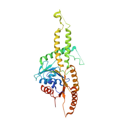
Model for eukaryotic tail-anchored protein binding based on the structure of Get3

Suloway, C.J.Chartron, J.W.Zaslaver, M.Clemons, W.M.

(2009) Proc Natl Acad Sci U S A 106: 14849-14854

  • DOI: https://doi.org/10.1073/pnas.0907522106
  • Primary Citation Related Structures: 
    3IBG, 3IDQ

  • PubMed Abstract: 

    The Get3 ATPase directs the delivery of tail-anchored (TA) proteins to the endoplasmic reticulum (ER). TA-proteins are characterized by having a single transmembrane helix (TM) at their extreme C terminus and include many essential proteins, such as SNAREs, apoptosis factors, and protein translocation components. These proteins cannot follow the SRP-dependent co-translational pathway that typifies most integral membrane proteins; instead, post-translationally, these proteins are recognized and bound by Get3 then delivered to the ER in the ATP dependent Get pathway. To elucidate a molecular mechanism for TA protein binding by Get3 we have determined three crystal structures in apo and ADP forms from Saccharomyces cerevisae (ScGet3-apo) and Aspergillus fumigatus (AfGet3-apo and AfGet3-ADP). Using structural information, we generated mutants to confirm important interfaces and essential residues. These results point to a model of how Get3 couples ATP hydrolysis to the binding and release of TA-proteins.


  • Organizational Affiliation
    • Division of Chemistry and Chemical Engineering, California Institute of Technology, 1200 East California Boulevard, Pasadena, CA 91125, USA.

Macromolecule Content 

  • Total Structure Weight: 233.67 kDa 
  • Atom Count: 14,238 
  • Modeled Residue Count: 1,800 
  • Deposited Residue Count: 2,088 
  • Unique protein chains: 1

Macromolecules

Find similar proteins by:|  3D Structure
Entity ID: 1
MoleculeChains  Sequence LengthOrganismDetailsImage
ATPase, subunit of the Get complex
A, B, C, D, E
A, B, C, D, E, F
348Aspergillus fumigatusMutation(s): 0 
Gene Names: AFUA_3G11350
EC: 3.6.3.16 (PDB Primary Data), 3.6 (UniProt)
UniProt
Find proteins for Q4WY07 (Aspergillus fumigatus (strain ATCC MYA-4609 / CBS 101355 / FGSC A1100 / Af293))
Explore Q4WY07 
Go to UniProtKB:  Q4WY07
Entity Groups
Sequence Clusters30% Identity50% Identity70% Identity90% Identity95% Identity100% Identity
UniProt GroupQ4WY07
Sequence Annotations
Expand
Reference Sequence

Experimental Data & Validation

Experimental Data

  • Method: X-RAY DIFFRACTION
  • Resolution: 3.20 Å
  • R-Value Free:  0.251 (Depositor), 0.244 (DCC) 
  • R-Value Work:  0.212 (Depositor), 0.205 (DCC) 
  • R-Value Observed: 0.214 (Depositor) 
Space Group: P 21 21 21
Unit Cell:
Length ( Å )Angle ( ˚ )
a = 67.76α = 90
b = 154.78β = 90
c = 242.88γ = 90
Software Package:
Software NamePurpose
SCALAdata processing
SOLVEphasing
PHENIXrefinement
PDB_EXTRACTdata extraction
Blu-Icedata collection
MOSFLMdata reduction
SCALAdata scaling

Structure Validation

View Full Validation Report


Ligand Structure Quality Assessment 


Entry History 

Deposition Data

Revision History  (Full details and data files)

  • Version 1.0: 2009-08-25
    Type: Initial release
  • Version 1.1: 2011-07-13
    Changes: Version format compliance
  • Version 1.2: 2024-11-20
    Changes: Data collection, Database references, Derived calculations, Structure summary