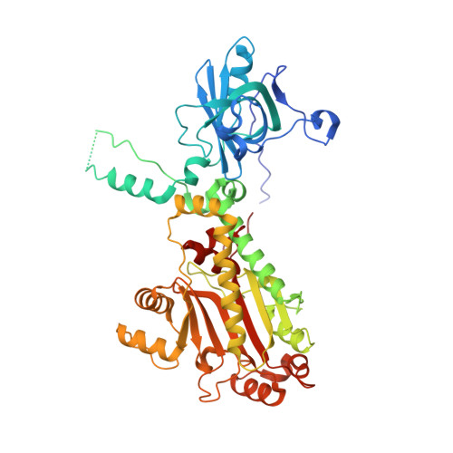
3I7F | pdb_00003i7f

Aspartyl tRNA synthetase from Entamoeba histolytica


Experimental Data Snapshot

  • Method: X-RAY DIFFRACTION
  • Resolution: 2.80 Å
  • R-Value Free: 
    0.237 (Depositor), 0.278 (DCC) 
  • R-Value Work: 
    0.200 (Depositor) 
  • R-Value Observed: 
    0.202 (Depositor) 

wwPDB Validation 3D Report Full Report

Validation slider image for 3I7F

This is version 1.3 of the entry. See complete history. 

Literature

Crystal structure of the aspartyl-tRNA synthetase from Entamoeba histolytica.

Merritt, E.A., Arakaki, T.L., Larson, E.T., Kelley, A., Mueller, N., Napuli, A.J., Zhang, L., Deditta, G., Luft, J., Verlinde, C.L., Fan, E., Zucker, F., Buckner, F.S., Van Voorhis, W.C., Hol, W.G.

(2010) Mol Biochem Parasitol 169: 95-100

  • DOI: https://doi.org/10.1016/j.molbiopara.2009.10.005
  • Primary Citation Related Structures: 
    3I7F

  • PubMed Abstract: 

    The crystal structure of the aspartyl-tRNA synthetase from the eukaryotic parasite Entamoeba histolytica has been determined at 2.8Aresolution. Relative to homologous sequences, the E. histolytica protein contains a 43-residue insertion between the N-terminal anticodon binding domain and the C-terminal catalytic domain. The present structure reveals that this insertion extends an arm of the hinge region that has previously been shown to mediate interaction of aspartyl-tRNA synthetase with the cognate tRNA D-stem. Modeling indicates that this Entamoeba-specific insertion is likely to increase the interaction surface with the cognate tRNA(Asp). In doing so it may substitute functionally for an RNA-binding motif located in N-terminal extensions found in AspRS sequences from lower eukaryotes but absent in Entamoeba. The E. histolytica AspRS structure shows a well-ordered N-terminus that contributes to the AspRS dimer interface.


  • Organizational Affiliation: 
    • Department of Biochemistry, University of Washington, Mailstop 357742, Seattle, WA 98195, USA. merritt@u.washington.edu

Macromolecule Content 

  • Total Structure Weight: 125.95 kDa 
  • Atom Count: 7,737 
  • Modeled Residue Count: 968 
  • Deposited Residue Count: 1,096 
  • Unique protein chains: 1

Macromolecules

Find similar proteins by:|  3D Structure
Entity ID: 1
MoleculeChains  Sequence LengthOrganismDetailsImage
Aspartyl-tRNA synthetase
A, B
548Entamoeba histolyticaMutation(s): 0 
Gene Names: EHI_175050
EC: 6.1.1.12
UniProt
Find proteins for C4LZN0 (Entamoeba histolytica (strain ATCC 30459 / HM-1:IMSS / ABRM))
Explore C4LZN0 
Go to UniProtKB:  C4LZN0
Entity Groups
Sequence Clusters30% Identity50% Identity70% Identity90% Identity95% Identity100% Identity
UniProt GroupC4LZN0
Sequence Annotations
Expand
Reference Sequence

Experimental Data & Validation

Experimental Data

  • Method: X-RAY DIFFRACTION
  • Resolution: 2.80 Å
  • R-Value Free:  0.237 (Depositor), 0.278 (DCC) 
  • R-Value Work:  0.200 (Depositor) 
  • R-Value Observed: 0.202 (Depositor) 
Space Group: H 3
Unit Cell:
Length ( Å )Angle ( ˚ )
a = 247.848α = 90
b = 247.848β = 90
c = 56.295γ = 120
Software Package:
Software NamePurpose
DENZOdata reduction
SCALEPACKdata scaling
REFMACrefinement
PDB_EXTRACTdata extraction
Blu-Icedata collection
BALBESphasing

Structure Validation

View Full Validation Report


Entry History 

Deposition Data

Revision History  (Full details and data files)

  • Version 1.0: 2009-07-21
    Type: Initial release
  • Version 1.1: 2011-07-13
    Changes: Advisory, Version format compliance
  • Version 1.2: 2017-11-01
    Changes: Refinement description
  • Version 1.3: 2024-02-21
    Changes: Data collection, Database references, Refinement description