3I6O | pdb_00003i6o

Crystal structure of wild type HIV-1 protease with macrocyclic inhibitor GRL-0216A


Experimental Data Snapshot

  • Method: X-RAY DIFFRACTION
  • Resolution: 1.17 Å
  • R-Value Free: 
    0.196 (Depositor), 0.195 (DCC) 
  • R-Value Work: 
    0.159 (Depositor) 
  • R-Value Observed: 
    0.161 (Depositor) 

Starting Model: experimental
View more details

wwPDB Validation 3D Report Full Report

Validation slider image for 3I6O

Ligand Structure Quality Assessment 


This is version 1.4 of the entry. See complete history

Literature

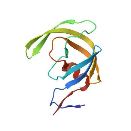
Design, Synthesis, Protein-Ligand X-ray Structure, and Biological Evaluation of a Series of Novel Macrocyclic Human Immunodeficiency Virus-1 Protease Inhibitors to Combat Drug Resistance.

Ghosh, A.K.Kulkarni, S.Anderson, D.D.Hong, L.Baldridge, A.Wang, Y.F.Chumanevich, A.A.Kovalevsky, A.Y.Tojo, Y.Amano, M.Koh, Y.Tang, J.Weber, I.T.Mitsuya, H.

(2009) J Med Chem 52: 7689-7705

  • DOI: https://doi.org/10.1021/jm900695w
  • Primary Citation Related Structures: 
    3I6O, 3I7E

  • PubMed Abstract: 

    The structure-based design, synthesis, and biological evaluation of a series of nonpeptidic macrocyclic HIV protease inhibitors are described. The inhibitors are designed to effectively fill in the hydrophobic pocket in the S1'-S2' subsites and retain all major hydrogen bonding interactions with the protein backbone similar to darunavir (1) or inhibitor 2. The ring size, the effect of methyl substitution, and unsaturation within the macrocyclic ring structure were assessed. In general, cyclic inhibitors were significantly more potent than their acyclic homologues, saturated rings were less active than their unsaturated analogues and a preference for 10- and 13-membered macrocylic rings was revealed. The addition of methyl substituents resulted in a reduction of potency. Both inhibitors 14b and 14c exhibited marked enzyme inhibitory and antiviral activity, and they exerted potent activity against multidrug-resistant HIV-1 variants. Protein-ligand X-ray structures of inhibitors 2 and 14c provided critical molecular insights into the ligand-binding site interactions.


  • Organizational Affiliation
    • Department of Chemistry and Medicinal Chemistry, Purdue University, West Lafayette, Indiana 47907, USA. akghosh@purdue.edu

Macromolecule Content 

  • Total Structure Weight: 24.75 kDa 
  • Atom Count: 1,972 
  • Modeled Residue Count: 198 
  • Deposited Residue Count: 198 
  • Unique protein chains: 1

Macromolecules

Find similar proteins by:|  3D Structure
Entity ID: 1
MoleculeChains  Sequence LengthOrganismDetailsImage
Protease
A, B
99Human immunodeficiency virus type 1 (BRU ISOLATE)Mutation(s): 5 
Gene Names: gag-pol
EC: 3.4.23.16
UniProt
Find proteins for P03367 (Human immunodeficiency virus type 1 group M subtype B (isolate BRU/LAI))
Explore P03367 
Go to UniProtKB:  P03367
Entity Groups
Sequence Clusters30% Identity50% Identity70% Identity90% Identity95% Identity100% Identity
UniProt GroupP03367
Sequence Annotations
Expand
Reference Sequence

Small Molecules

Ligands 4 Unique
IDChains Name / Formula / InChI Key2D Diagram3D Interactions
GR6

Query on GR6


Download:Ideal Coordinates CCD File
Y [auth B](3R,3aS,6aR)-hexahydrofuro[2,3-b]furan-3-yl {(1S,2R)-1-benzyl-2-hydroxy-3-[(7E)-13-methoxy-1,1-dioxido-3,4,5,6,9,10-hexahydro-2H-11,1,2-benzoxathiazacyclotridecin-2-yl]propyl}carbamate
C32 H42 N2 O9 S
ZQJBDIZBNGQJDT-JZAADHNPSA-N
IOD

Query on IOD


Download:Ideal Coordinates CCD File
D [auth A]
E [auth A]
F [auth A]
G [auth A]
H [auth A]
D [auth A],
E [auth A],
F [auth A],
G [auth A],
H [auth A],
I [auth A],
J [auth A],
K [auth A],
N [auth B],
O [auth B],
P [auth B],
Q [auth B],
R [auth B],
S [auth B],
T [auth B],
U [auth B],
V [auth B],
W [auth B],
X [auth B]
IODIDE ION
I
XMBWDFGMSWQBCA-UHFFFAOYSA-M
GOL

Query on GOL


Download:Ideal Coordinates CCD File
L [auth A],
Z [auth B]
GLYCEROL
C3 H8 O3
PEDCQBHIVMGVHV-UHFFFAOYSA-N
NA

Query on NA


Download:Ideal Coordinates CCD File
C [auth A],
M [auth B]
SODIUM ION
Na
FKNQFGJONOIPTF-UHFFFAOYSA-N

Experimental Data & Validation

Experimental Data

  • Method: X-RAY DIFFRACTION
  • Resolution: 1.17 Å
  • R-Value Free:  0.196 (Depositor), 0.195 (DCC) 
  • R-Value Work:  0.159 (Depositor) 
  • R-Value Observed: 0.161 (Depositor) 
Space Group: P 21 21 2
Unit Cell:
Length ( Å )Angle ( ˚ )
a = 58.764α = 90
b = 86.328β = 90
c = 45.976γ = 90
Software Package:
Software NamePurpose
MAR345data collection
PHASERphasing
SHELXL-97refinement
HKL-2000data reduction
HKL-2000data scaling

Structure Validation

View Full Validation Report


Ligand Structure Quality Assessment 


Entry History 

Deposition Data

Revision History  (Full details and data files)

  • Version 1.0: 2009-09-29
    Type: Initial release
  • Version 1.1: 2011-07-13
    Changes: Non-polymer description, Version format compliance
  • Version 1.2: 2017-11-01
    Changes: Refinement description
  • Version 1.3: 2021-10-13
    Changes: Database references, Derived calculations
  • Version 1.4: 2023-09-06
    Changes: Data collection, Refinement description