3I1C | pdb_00003i1c

Crystal Structure of a Novel Engineered Diels-Alderase: DA_20_00_A74I


Experimental Data Snapshot

  • Method: X-RAY DIFFRACTION
  • Resolution: 2.20 Å
  • R-Value Free: 
    0.253 (Depositor), 0.268 (DCC) 
  • R-Value Work: 
    0.184 (Depositor), 0.196 (DCC) 
  • R-Value Observed: 
    0.188 (Depositor) 

wwPDB Validation 3D Report Full Report

Validation slider image for 3I1C

This is version 1.3 of the entry. See complete history

Literature

Computational design of an enzyme catalyst for a stereoselective bimolecular Diels-Alder reaction.

Siegel, J.B.Zanghellini, A.Lovick, H.M.Kiss, G.Lambert, A.R.St Clair, J.L.Gallaher, J.L.Hilvert, D.Gelb, M.H.Stoddard, B.L.Houk, K.N.Michael, F.E.Baker, D.

(2010) Science 329: 309-313

  • DOI: https://doi.org/10.1126/science.1190239
  • Primary Citation Related Structures: 
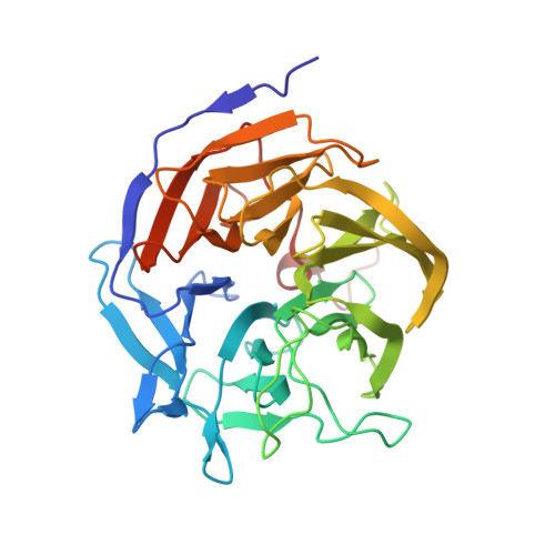
    3I1C

  • PubMed Abstract: 

    The Diels-Alder reaction is a cornerstone in organic synthesis, forming two carbon-carbon bonds and up to four new stereogenic centers in one step. No naturally occurring enzymes have been shown to catalyze bimolecular Diels-Alder reactions. We describe the de novo computational design and experimental characterization of enzymes catalyzing a bimolecular Diels-Alder reaction with high stereoselectivity and substrate specificity. X-ray crystallography confirms that the structure matches the design for the most active of the enzymes, and binding site substitutions reprogram the substrate specificity. Designed stereoselective catalysts for carbon-carbon bond-forming reactions should be broadly useful in synthetic chemistry.


  • Organizational Affiliation
    • Department of Biochemistry, University of Washington, Seattle, WA 98195, USA.

Macromolecule Content 

  • Total Structure Weight: 36.19 kDa 
  • Atom Count: 2,787 
  • Modeled Residue Count: 314 
  • Deposited Residue Count: 324 
  • Unique protein chains: 1

Macromolecules

Find similar proteins by:|  3D Structure
Entity ID: 1
MoleculeChains  Sequence LengthOrganismDetailsImage
Diisopropyl-fluorophosphatase324Loligo vulgarisMutation(s): 14 
EC: 3.1.8.2 (PDB Primary Data), 3.8.2.2 (UniProt)
UniProt
Find proteins for Q7SIG4 (Loligo vulgaris)
Explore Q7SIG4 
Go to UniProtKB:  Q7SIG4
Entity Groups
Sequence Clusters30% Identity50% Identity70% Identity90% Identity95% Identity100% Identity
UniProt GroupQ7SIG4
Sequence Annotations
Expand
Reference Sequence

Small Molecules

Ligands 1 Unique
IDChains Name / Formula / InChI Key2D Diagram3D Interactions
GOL

Query on GOL


Download:Ideal Coordinates CCD File
B [auth A]GLYCEROL
C3 H8 O3
PEDCQBHIVMGVHV-UHFFFAOYSA-N

Experimental Data & Validation

Experimental Data

  • Method: X-RAY DIFFRACTION
  • Resolution: 2.20 Å
  • R-Value Free:  0.253 (Depositor), 0.268 (DCC) 
  • R-Value Work:  0.184 (Depositor), 0.196 (DCC) 
  • R-Value Observed: 0.188 (Depositor) 
Space Group: P 21 21 21
Unit Cell:
Length ( Å )Angle ( ˚ )
a = 41.239α = 90
b = 83.112β = 90
c = 85.18γ = 90
Software Package:
Software NamePurpose
REFMACrefinement

Structure Validation

View Full Validation Report


Entry History 

Deposition Data

Revision History  (Full details and data files)

  • Version 1.0: 2009-08-18
    Type: Initial release
  • Version 1.1: 2011-07-13
    Changes: Non-polymer description, Version format compliance
  • Version 1.2: 2021-10-13
    Changes: Database references, Derived calculations
  • Version 1.3: 2024-02-21
    Changes: Data collection