


Crystal structure of Periplasmic Binding Protein BMA293 (YP_104442.1) from Burkholderia mallei ATCC 23344 at 1.80 A resolution
Joint Center for Structural Genomics (JCSG)To be published.
Experimental Data Snapshot
wwPDB Validation 3D Report Full Report

Entity ID: 1 | |||||
|---|---|---|---|---|---|
| Molecule | Chains | Sequence Length | Organism | Details | Image |
| Periplasmic branched-chain amino acid-binding protein | 375 | Burkholderia mallei | Mutation(s): 0 Gene Names: BMA2936, YP_104442.1 | 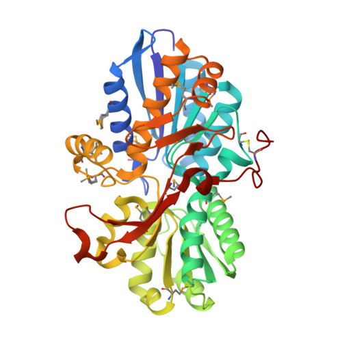 | |
UniProt | |||||
Find proteins for A0A0H2WF95 (Burkholderia mallei (strain ATCC 23344)) Explore A0A0H2WF95 Go to UniProtKB: A0A0H2WF95 | |||||
Entity Groups | |||||
| Sequence Clusters | 30% Identity50% Identity70% Identity90% Identity95% Identity100% Identity | ||||
| UniProt Group | A0A0H2WF95 | ||||
Sequence AnnotationsExpand | |||||
Reference Sequence | |||||
| Ligands 3 Unique | |||||
|---|---|---|---|---|---|
| ID | Chains | Name / Formula / InChI Key | 2D Diagram | 3D Interactions | |
| CIT Download:Ideal Coordinates CCD File | D [auth A] E [auth A] F [auth A] P [auth B] Q [auth B] | CITRIC ACID C6 H8 O7 KRKNYBCHXYNGOX-UHFFFAOYSA-N |  | ||
| AAE Download:Ideal Coordinates CCD File | C [auth A], O [auth B] | ACETOACETIC ACID C4 H6 O3 WDJHALXBUFZDSR-UHFFFAOYSA-N |  | ||
| EDO Download:Ideal Coordinates CCD File | G [auth A] H [auth A] I [auth A] J [auth A] K [auth A] | 1,2-ETHANEDIOL C2 H6 O2 LYCAIKOWRPUZTN-UHFFFAOYSA-N |  | ||
| Modified Residues 1 Unique | |||||
|---|---|---|---|---|---|
| ID | Chains | Type | Formula | 2D Diagram | Parent |
| MSE Query on MSE | A, B | L-PEPTIDE LINKING | C5 H11 N O2 Se |  | MET |
| Length ( Å ) | Angle ( ˚ ) |
|---|---|
| a = 58.255 | α = 90 |
| b = 81.151 | β = 92.86 |
| c = 73.996 | γ = 90 |
| Software Name | Purpose |
|---|---|
| REFMAC | refinement |
| PHENIX | refinement |
| SHELX | phasing |
| MolProbity | model building |
| XSCALE | data scaling |
| PDB_EXTRACT | data extraction |
| XDS | data reduction |
| SHELXD | phasing |
| autoSHARP | phasing |


