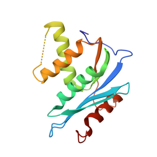
3HYF

Crystal structure of HIV-1 RNase H p15 with engineered E. coli loop and active site inhibitor


Experimental Data Snapshot

  • Method: X-RAY DIFFRACTION
  • Resolution: 1.70 Å
  • R-Value Free: 0.232 
  • R-Value Work: 0.196 
  • R-Value Observed: 0.198 

Starting Model: experimental
View more details

wwPDB Validation   3D Report Full Report


Ligand Structure Quality Assessment 


This is version 1.5 of the entry. See complete history


Literature

RNase H active site inhibitors of human immunodeficiency virus type 1 reverse transcriptase: design, biochemical activity, and structural information.

Kirschberg, T.A.Balakrishnan, M.Squires, N.H.Barnes, T.Brendza, K.M.Chen, X.Eisenberg, E.J.Jin, W.Kutty, N.Leavitt, S.Liclican, A.Liu, Q.Liu, X.Mak, J.Perry, J.K.Wang, M.Watkins, W.J.Lansdon, E.B.

(2009) J Med Chem 52: 5781-5784

  • DOI: https://doi.org/10.1021/jm900597q
  • Primary Citation of Related Structures:  
    3HYF

  • PubMed Abstract: 

    Pyrimidinol carboxylic acids were designed as inhibitors of HIV-1 RNase H function. These molecules can coordinate to two divalent metal ions in the RNase H active site. Inhibition of enzymatic activity was measured in a biochemical assay, but no antiviral effect was observed. Binding was demonstrated via a solid state structure of the isolated p15-Ec domain of HIV-1 RT showing inhibitor and two Mn(II) ions bound to the RNase H active site.


  • Organizational Affiliation

    Department of Medicinal Chemistry, Gilead Sciences, Foster City, California 94404, USA. tkirschberg@gilead.com


Macromolecules
Find similar proteins by:  (by identity cutoff)  |  3D Structure
Entity ID: 1
MoleculeChains Sequence LengthOrganismDetailsImage
Reverse transcriptase/RNaseH150Human immunodeficiency virus 1Escherichia coli K-12Mutation(s): 0 
Gene Names: polReverse Transcriptase (amino acids 427-560)
EC: 3.1.26.4
UniProt
Find proteins for P0A7Y4 (Escherichia coli (strain K12))
Explore P0A7Y4 
Go to UniProtKB:  P0A7Y4
Find proteins for Q72547 (Human immunodeficiency virus 1)
Explore Q72547 
Go to UniProtKB:  Q72547
Entity Groups  
Sequence Clusters30% Identity50% Identity70% Identity90% Identity95% Identity100% Identity
UniProt GroupsQ72547P0A7Y4
Sequence Annotations
Expand
  • Reference Sequence
Small Molecules
Ligands 4 Unique
IDChains Name / Formula / InChI Key2D Diagram3D Interactions
ON1
Query on ON1

Download Ideal Coordinates CCD File 
G [auth A]2-(3,4-dichlorobenzyl)-5,6-dihydroxypyrimidine-4-carboxylic acid
C12 H8 Cl2 N2 O4
OZDSTVKQUHMIRA-UHFFFAOYSA-N
GOL
Query on GOL

Download Ideal Coordinates CCD File 
E [auth A],
F [auth A]
GLYCEROL
C3 H8 O3
PEDCQBHIVMGVHV-UHFFFAOYSA-N
ACT
Query on ACT

Download Ideal Coordinates CCD File 
D [auth A]ACETATE ION
C2 H3 O2
QTBSBXVTEAMEQO-UHFFFAOYSA-M
MN
Query on MN

Download Ideal Coordinates CCD File 
B [auth A],
C [auth A]
MANGANESE (II) ION
Mn
WAEMQWOKJMHJLA-UHFFFAOYSA-N
Binding Affinity Annotations 
IDSourceBinding Affinity
ON1 PDBBind:  3HYF IC50: 1200 (nM) from 1 assay(s)
Experimental Data & Validation

Experimental Data

  • Method: X-RAY DIFFRACTION
  • Resolution: 1.70 Å
  • R-Value Free: 0.232 
  • R-Value Work: 0.196 
  • R-Value Observed: 0.198 
  • Space Group: I 21 21 21
Unit Cell:
Length ( Å )Angle ( ˚ )
a = 39.717α = 90
b = 90.326β = 90
c = 113.334γ = 90
Software Package:
Software NamePurpose
BOSdata collection
AMoREphasing
CNXrefinement
HKL-2000data reduction
HKL-2000data scaling

Structure Validation

View Full Validation Report


Ligand Structure Quality Assessment 


Entry History 

Deposition Data

Revision History  (Full details and data files)

  • Version 1.0: 2009-10-20
    Type: Initial release
  • Version 1.1: 2011-07-13
    Changes: Non-polymer description, Version format compliance
  • Version 1.2: 2011-11-16
    Changes: Atomic model
  • Version 1.3: 2017-08-16
    Changes: Refinement description, Source and taxonomy
  • Version 1.4: 2023-09-06
    Changes: Data collection, Database references, Derived calculations, Refinement description
  • Version 1.5: 2024-03-13
    Changes: Source and taxonomy