Unusual Diheme Conformation of the Heme-Degrading Protein from Mycobacterium tuberculosis
Chim, N., Iniguez, A., Nguyen, T.Q., Goulding, C.W.(2009) J Mol Biology 395: 595-608
- PubMed: 19917297 Search on PubMedSearch on PubMed Central
- DOI: https://doi.org/10.1016/j.jmb.2009.11.025
- Primary Citation Related Structures:
3HX9 - PubMed Abstract:
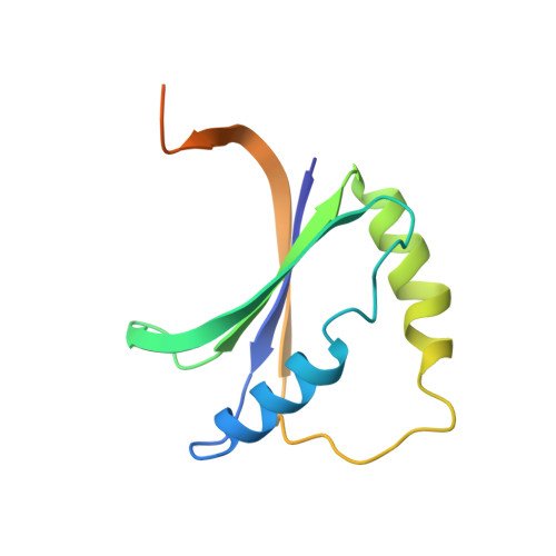
Heme degradation plays a pivotal role in the availability of the essential nutrient, iron, in pathogenic bacteria. A previously unannotated protein from Mycobacterium tuberculosis, Rv3592, which shares homology to heme-degrading enzymes, has been identified. Biochemical analyses confirm that Rv3592, which we have termed MhuD (mycobacterial heme utilization, degrader), is able to bind and degrade heme. Interestingly, contrary to previously reported stoichiometry for the Staphylococcus aureus heme degraders, iron-regulated surface determinant (Isd)G and IsdI, MhuD has the ability to bind heme in a 1:2 protein-to-heme ratio, although the MhuD-diheme complex is inactive. Furthermore, the 1.75-A crystal structure of the MhuD-diheme complex reveals two stacked hemes forming extensive contacts with residues in the active site. In particular, the solvent-exposed heme is axially liganded by His75 and is stacked planar upon the solvent-protected heme. The solvent-protected heme is coordinated by a chloride ion, which is, in turn, stabilized by Asn7. Structural comparison between MhuD-diheme and inactive IsdG and IsdI bound to only one highly distorted metalloporphyrin ring reveals that several residues located in alpha-helix 2 and the subsequent loop appear to be responsible for heme stoichiometric differences and suggest open and closed conformations for substrate entry and product exit.
- Department of Molecular Biology and Biochemistry, University of California, Irvine, CA 92697, USA.
Organizational Affiliation: